Controlled Experiment
Saul McLeod, PhD
Editor-in-Chief for Simply Psychology
BSc (Hons) Psychology, MRes, PhD, University of Manchester
Saul McLeod, PhD., is a qualified psychology teacher with over 18 years of experience in further and higher education. He has been published in peer-reviewed journals, including the Journal of Clinical Psychology.
Learn about our Editorial Process
Olivia Guy-Evans, MSc
Associate Editor for Simply Psychology
BSc (Hons) Psychology, MSc Psychology of Education
Olivia Guy-Evans is a writer and associate editor for Simply Psychology. She has previously worked in healthcare and educational sectors.
This is when a hypothesis is scientifically tested.
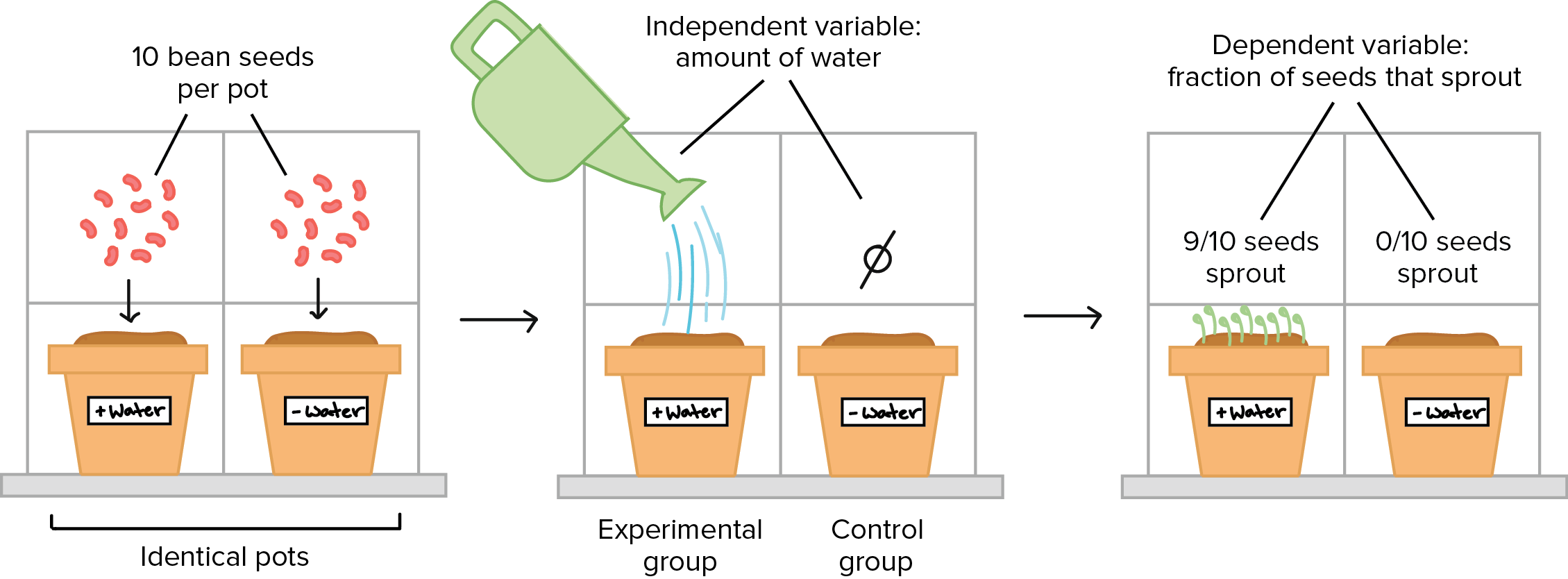
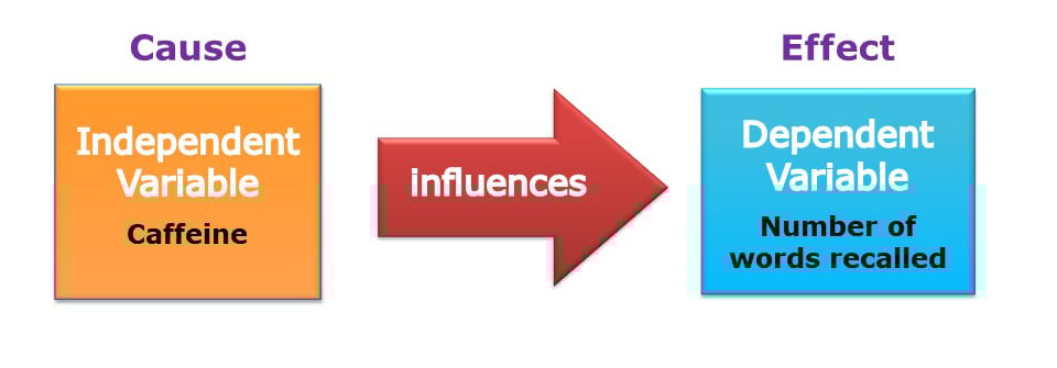
In a controlled experiment, an independent variable (the cause) is systematically manipulated, and the dependent variable (the effect) is measured; any extraneous variables are controlled.
The researcher can operationalize (i.e., define) the studied variables so they can be objectively measured. The quantitative data can be analyzed to see if there is a difference between the experimental and control groups.
What is the control group?
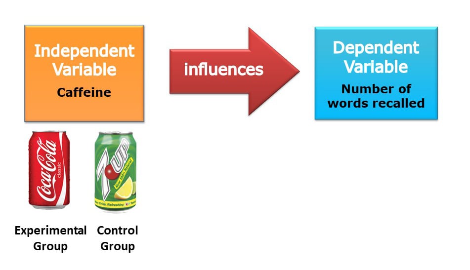
In experiments scientists compare a control group and an experimental group that are identical in all respects, except for one difference – experimental manipulation.
Unlike the experimental group, the control group is not exposed to the independent variable under investigation and so provides a baseline against which any changes in the experimental group can be compared.
Since experimental manipulation is the only difference between the experimental and control groups, we can be sure that any differences between the two are due to experimental manipulation rather than chance.
Randomly allocating participants to independent variable groups means that all participants should have an equal chance of participating in each condition.
The principle of random allocation is to avoid bias in how the experiment is carried out and limit the effects of participant variables.
What are extraneous variables?
The researcher wants to ensure that the manipulation of the independent variable has changed the changes in the dependent variable.
Hence, all the other variables that could affect the dependent variable to change must be controlled. These other variables are called extraneous or confounding variables.
Extraneous variables should be controlled were possible, as they might be important enough to provide alternative explanations for the effects.
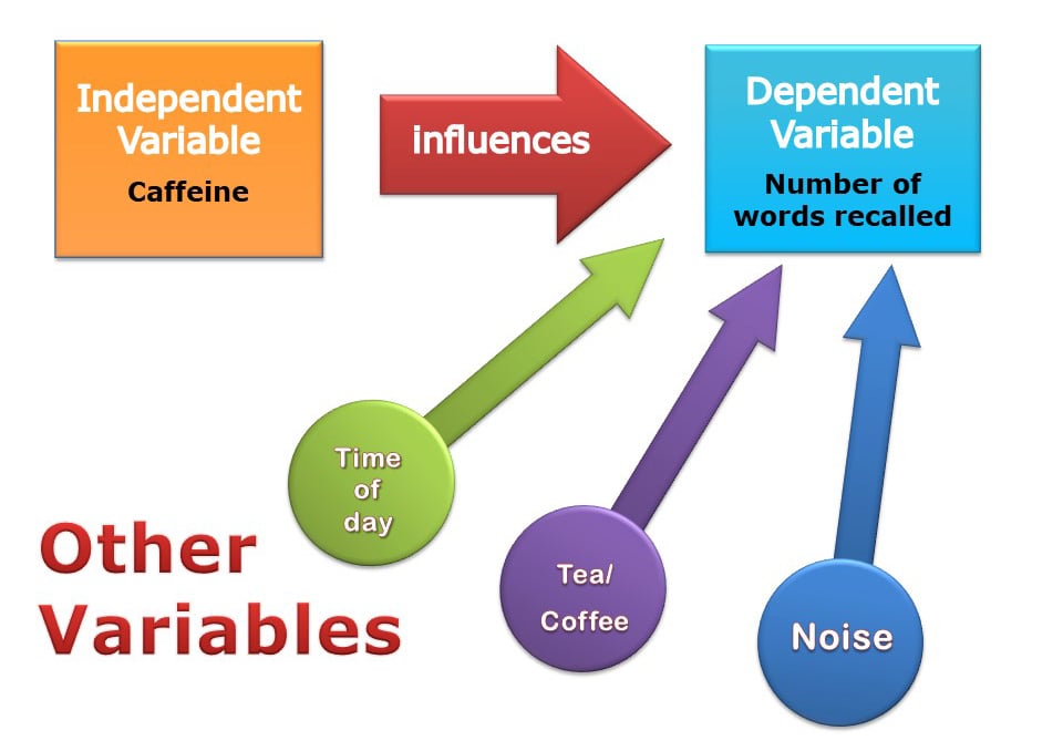
In practice, it would be difficult to control all the variables in a child’s educational achievement. For example, it would be difficult to control variables that have happened in the past.
A researcher can only control the current environment of participants, such as time of day and noise levels.
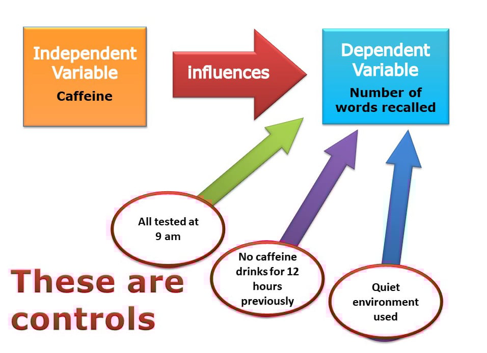
Why conduct controlled experiments?
Scientists use controlled experiments because they allow for precise control of extraneous and independent variables. This allows a cause-and-effect relationship to be established.
Controlled experiments also follow a standardized step-by-step procedure. This makes it easy for another researcher to replicate the study.
Key Terminology
Experimental group.
The group being treated or otherwise manipulated for the sake of the experiment.
Control Group
They receive no treatment and are used as a comparison group.
Ecological validity
The degree to which an investigation represents real-life experiences.
Experimenter effects
These are the ways that the experimenter can accidentally influence the participant through their appearance or behavior.
Demand characteristics
The clues in an experiment lead the participants to think they know what the researcher is looking for (e.g., the experimenter’s body language).
Independent variable (IV)
The variable the experimenter manipulates (i.e., changes) – is assumed to have a direct effect on the dependent variable.
Dependent variable (DV)
Variable the experimenter measures. This is the outcome (i.e., the result) of a study.
Extraneous variables (EV)
All variables that are not independent variables but could affect the results (DV) of the experiment. Extraneous variables should be controlled where possible.
Confounding variables
Variable(s) that have affected the results (DV), apart from the IV. A confounding variable could be an extraneous variable that has not been controlled.
Random Allocation
Randomly allocating participants to independent variable conditions means that all participants should have an equal chance of participating in each condition.
Order effects
Changes in participants’ performance due to their repeating the same or similar test more than once. Examples of order effects include:
(i) practice effect: an improvement in performance on a task due to repetition, for example, because of familiarity with the task;
(ii) fatigue effect: a decrease in performance of a task due to repetition, for example, because of boredom or tiredness.
What is the control in an experiment?
In an experiment , the control is a standard or baseline group not exposed to the experimental treatment or manipulation. It serves as a comparison group to the experimental group, which does receive the treatment or manipulation.
The control group helps to account for other variables that might influence the outcome, allowing researchers to attribute differences in results more confidently to the experimental treatment.
Establishing a cause-and-effect relationship between the manipulated variable (independent variable) and the outcome (dependent variable) is critical in establishing a cause-and-effect relationship between the manipulated variable.
What is the purpose of controlling the environment when testing a hypothesis?
Controlling the environment when testing a hypothesis aims to eliminate or minimize the influence of extraneous variables. These variables other than the independent variable might affect the dependent variable, potentially confounding the results.
By controlling the environment, researchers can ensure that any observed changes in the dependent variable are likely due to the manipulation of the independent variable, not other factors.
This enhances the experiment’s validity, allowing for more accurate conclusions about cause-and-effect relationships.
It also improves the experiment’s replicability, meaning other researchers can repeat the experiment under the same conditions to verify the results.
Why are hypotheses important to controlled experiments?
Hypotheses are crucial to controlled experiments because they provide a clear focus and direction for the research. A hypothesis is a testable prediction about the relationship between variables.
It guides the design of the experiment, including what variables to manipulate (independent variables) and what outcomes to measure (dependent variables).
The experiment is then conducted to test the validity of the hypothesis. If the results align with the hypothesis, they provide evidence supporting it.
The hypothesis may be revised or rejected if the results do not align. Thus, hypotheses are central to the scientific method, driving the iterative inquiry, experimentation, and knowledge advancement process.
What is the experimental method?
The experimental method is a systematic approach in scientific research where an independent variable is manipulated to observe its effect on a dependent variable, under controlled conditions.
Have a language expert improve your writing
Run a free plagiarism check in 10 minutes, automatically generate references for free.
- Knowledge Base
- Methodology
- Controlled Experiments | Methods & Examples of Control
Controlled Experiments | Methods & Examples of Control
Published on 19 April 2022 by Pritha Bhandari . Revised on 10 October 2022.
In experiments , researchers manipulate independent variables to test their effects on dependent variables. In a controlled experiment , all variables other than the independent variable are controlled or held constant so they don’t influence the dependent variable.
Controlling variables can involve:
- Holding variables at a constant or restricted level (e.g., keeping room temperature fixed)
- Measuring variables to statistically control for them in your analyses
- Balancing variables across your experiment through randomisation (e.g., using a random order of tasks)
Table of contents
Why does control matter in experiments, methods of control, problems with controlled experiments, frequently asked questions about controlled experiments.
Control in experiments is critical for internal validity , which allows you to establish a cause-and-effect relationship between variables.
- Your independent variable is the colour used in advertising.
- Your dependent variable is the price that participants are willing to pay for a standard fast food meal.
Extraneous variables are factors that you’re not interested in studying, but that can still influence the dependent variable. For strong internal validity, you need to remove their effects from your experiment.
- Design and description of the meal
- Study environment (e.g., temperature or lighting)
- Participant’s frequency of buying fast food
- Participant’s familiarity with the specific fast food brand
- Participant’s socioeconomic status
Prevent plagiarism, run a free check.
You can control some variables by standardising your data collection procedures. All participants should be tested in the same environment with identical materials. Only the independent variable (e.g., advert colour) should be systematically changed between groups.
Other extraneous variables can be controlled through your sampling procedures . Ideally, you’ll select a sample that’s representative of your target population by using relevant inclusion and exclusion criteria (e.g., including participants from a specific income bracket, and not including participants with colour blindness).
By measuring extraneous participant variables (e.g., age or gender) that may affect your experimental results, you can also include them in later analyses.
After gathering your participants, you’ll need to place them into groups to test different independent variable treatments. The types of groups and method of assigning participants to groups will help you implement control in your experiment.
Control groups
Controlled experiments require control groups . Control groups allow you to test a comparable treatment, no treatment, or a fake treatment, and compare the outcome with your experimental treatment.
You can assess whether it’s your treatment specifically that caused the outcomes, or whether time or any other treatment might have resulted in the same effects.
- A control group that’s presented with red advertisements for a fast food meal
- An experimental group that’s presented with green advertisements for the same fast food meal
Random assignment
To avoid systematic differences between the participants in your control and treatment groups, you should use random assignment .
This helps ensure that any extraneous participant variables are evenly distributed, allowing for a valid comparison between groups .
Random assignment is a hallmark of a ‘true experiment’ – it differentiates true experiments from quasi-experiments .
Masking (blinding)
Masking in experiments means hiding condition assignment from participants or researchers – or, in a double-blind study , from both. It’s often used in clinical studies that test new treatments or drugs.
Sometimes, researchers may unintentionally encourage participants to behave in ways that support their hypotheses. In other cases, cues in the study environment may signal the goal of the experiment to participants and influence their responses.
Using masking means that participants don’t know whether they’re in the control group or the experimental group. This helps you control biases from participants or researchers that could influence your study results.
Although controlled experiments are the strongest way to test causal relationships, they also involve some challenges.
Difficult to control all variables
Especially in research with human participants, it’s impossible to hold all extraneous variables constant, because every individual has different experiences that may influence their perception, attitudes, or behaviors.
But measuring or restricting extraneous variables allows you to limit their influence or statistically control for them in your study.
Risk of low external validity
Controlled experiments have disadvantages when it comes to external validity – the extent to which your results can be generalised to broad populations and settings.
The more controlled your experiment is, the less it resembles real world contexts. That makes it harder to apply your findings outside of a controlled setting.
There’s always a tradeoff between internal and external validity . It’s important to consider your research aims when deciding whether to prioritise control or generalisability in your experiment.
Experimental designs are a set of procedures that you plan in order to examine the relationship between variables that interest you.
To design a successful experiment, first identify:
- A testable hypothesis
- One or more independent variables that you will manipulate
- One or more dependent variables that you will measure
When designing the experiment, first decide:
- How your variable(s) will be manipulated
- How you will control for any potential confounding or lurking variables
- How many subjects you will include
- How you will assign treatments to your subjects
Cite this Scribbr article
If you want to cite this source, you can copy and paste the citation or click the ‘Cite this Scribbr article’ button to automatically add the citation to our free Reference Generator.
Bhandari, P. (2022, October 10). Controlled Experiments | Methods & Examples of Control. Scribbr. Retrieved 5 November 2024, from https://www.scribbr.co.uk/research-methods/controlled-experiments/
Is this article helpful?
Pritha Bhandari
LEARN STATISTICS EASILY
Learn Data Analysis Now!
What is: Controlled Experiment
What is a controlled experiment.
A controlled experiment is a scientific test that aims to establish a cause-and-effect relationship between variables. In this type of experiment, researchers manipulate one variable, known as the independent variable, while keeping all other variables constant. This allows for a clear observation of how changes in the independent variable affect the dependent variable, which is the outcome being measured. Controlled experiments are fundamental in fields such as statistics, data analysis , and data science, as they provide reliable data that can be analyzed to draw valid conclusions.
Ad description. Lorem ipsum dolor sit amet, consectetur adipiscing elit.
The Importance of Control Groups
In a controlled experiment, the use of control groups is essential. A control group is a baseline group that does not receive the experimental treatment or intervention. By comparing the results of the experimental group, which receives the treatment, to the control group, researchers can determine the effect of the independent variable more accurately. This comparison helps to eliminate alternative explanations for the observed effects, thereby strengthening the validity of the experiment’s conclusions.
Randomization in Controlled Experiments
Randomization is a critical aspect of controlled experiments. It involves randomly assigning participants or subjects to either the experimental group or the control group. This process helps to ensure that any differences observed between the groups are due to the treatment rather than pre-existing differences among the participants. Randomization minimizes bias and enhances the reliability of the results, making it a cornerstone of rigorous experimental design in data science.
Types of Controlled Experiments
Controlled experiments can be categorized into different types, including laboratory experiments and field experiments. Laboratory experiments are conducted in a controlled environment where researchers can manipulate variables with precision . In contrast, field experiments take place in natural settings, allowing researchers to observe real-world behaviors while still maintaining control over certain variables. Each type has its advantages and disadvantages, and the choice depends on the research question and context.
Hypothesis Testing in Controlled Experiments
Hypothesis testing is a fundamental component of controlled experiments. Researchers formulate a hypothesis, which is a testable prediction about the relationship between the independent and dependent variables. The controlled experiment is designed to test this hypothesis, and statistical methods are employed to analyze the data collected. If the results support the hypothesis, it may be accepted; if not, it may be rejected or revised. This process is crucial for advancing knowledge in statistics and data analysis.
Data Collection and Analysis
Data collection in controlled experiments is systematic and structured. Researchers gather quantitative or qualitative data based on the outcomes they are measuring. This data is then analyzed using various statistical techniques to determine the significance of the results. The analysis helps researchers understand the impact of the independent variable on the dependent variable, providing insights that can inform future research and decision-making in data science.
Ethical Considerations in Controlled Experiments
Ethical considerations are paramount in conducting controlled experiments, especially those involving human subjects. Researchers must ensure that participants are fully informed about the nature of the experiment and provide their consent. Additionally, they must consider the potential risks and benefits of the study. Adhering to ethical guidelines not only protects participants but also enhances the credibility of the research findings in the field of data analysis.
Limitations of Controlled Experiments
While controlled experiments are powerful tools for establishing causality, they do have limitations. One significant limitation is the artificial nature of laboratory settings, which may not accurately reflect real-world conditions. Additionally, controlled experiments may not always be feasible or ethical in certain situations, such as when studying complex social behaviors. Researchers must be aware of these limitations and consider complementary research methods to gain a comprehensive understanding of the phenomena being studied.
Applications of Controlled Experiments
Controlled experiments have wide-ranging applications across various fields, including psychology, medicine, and marketing. In psychology, they are used to study behavioral responses to different stimuli. In medicine, controlled clinical trials are essential for testing the efficacy of new treatments. In marketing, controlled experiments help businesses understand consumer preferences and optimize their strategies. The versatility of controlled experiments makes them invaluable in generating actionable insights from data analysis.
An official website of the United States government
Official websites use .gov A .gov website belongs to an official government organization in the United States.
Secure .gov websites use HTTPS A lock ( Lock Locked padlock icon ) or https:// means you've safely connected to the .gov website. Share sensitive information only on official, secure websites.
- Publications
- Account settings
- Advanced Search
- Journal List
Hypothesis tests
- Author information
- Article notes
- Copyright and License information
Accepted 2019 Mar 28; Issue date 2019 Jul.
Key points.
Hypothesis tests are used to assess whether a difference between two samples represents a real difference between the populations from which the samples were taken.
A null hypothesis of ‘no difference’ is taken as a starting point, and we calculate the probability that both sets of data came from the same population. This probability is expressed as a p -value.
When the null hypothesis is false, p- values tend to be small. When the null hypothesis is true, any p- value is equally likely.
Learning objectives.
By reading this article, you should be able to:
Explain why hypothesis testing is used.
Use a table to determine which hypothesis test should be used for a particular situation.
Interpret a p- value.
A hypothesis test is a procedure used in statistics to assess whether a particular viewpoint is likely to be true. They follow a strict protocol, and they generate a ‘ p- value’, on the basis of which a decision is made about the truth of the hypothesis under investigation. All of the routine statistical ‘tests’ used in research— t- tests, χ 2 tests, Mann–Whitney tests, etc.—are all hypothesis tests, and in spite of their differences they are all used in essentially the same way. But why do we use them at all?
Comparing the heights of two individuals is easy: we can measure their height in a standardised way and compare them. When we want to compare the heights of two small well-defined groups (for example two groups of children), we need to use a summary statistic that we can calculate for each group. Such summaries (means, medians, etc.) form the basis of descriptive statistics, and are well described elsewhere. 1 However, a problem arises when we try to compare very large groups or populations: it may be impractical or even impossible to take a measurement from everyone in the population, and by the time you do so, the population itself will have changed. A similar problem arises when we try to describe the effects of drugs—for example by how much on average does a particular vasopressor increase MAP?
To solve this problem, we use random samples to estimate values for populations. By convention, the values we calculate from samples are referred to as statistics and denoted by Latin letters ( x ¯ for sample mean; SD for sample standard deviation) while the unknown population values are called parameters , and denoted by Greek letters (μ for population mean, σ for population standard deviation).
Inferential statistics describes the methods we use to estimate population parameters from random samples; how we can quantify the level of inaccuracy in a sample statistic; and how we can go on to use these estimates to compare populations.
Sampling error
There are many reasons why a sample may give an inaccurate picture of the population it represents: it may be biased, it may not be big enough, and it may not be truly random. However, even if we have been careful to avoid these pitfalls, there is an inherent difference between the sample and the population at large. To illustrate this, let us imagine that the actual average height of males in London is 174 cm. If I were to sample 100 male Londoners and take a mean of their heights, I would be very unlikely to get exactly 174 cm. Furthermore, if somebody else were to perform the same exercise, it would be unlikely that they would get the same answer as I did. The sample mean is different each time it is taken, and the way it differs from the actual mean of the population is described by the standard error of the mean (standard error, or SEM ). The standard error is larger if there is a lot of variation in the population, and becomes smaller as the sample size increases. It is calculated thus:
where SD is the sample standard deviation, and n is the sample size.
As errors are normally distributed, we can use this to estimate a 95% confidence interval on our sample mean as follows:
We can interpret this as meaning ‘We are 95% confident that the actual mean is within this range.’
Some confusion arises at this point between the SD and the standard error. The SD is a measure of variation in the sample. The range x ¯ ± ( 1.96 × SD ) will normally contain 95% of all your data. It can be used to illustrate the spread of the data and shows what values are likely. In contrast, standard error tells you about the precision of the mean and is used to calculate confidence intervals.
One straightforward way to compare two samples is to use confidence intervals. If we calculate the mean height of two groups and find that the 95% confidence intervals do not overlap, this can be taken as evidence of a difference between the two means. This method of statistical inference is reasonably intuitive and can be used in many situations. 2 Many journals, however, prefer to report inferential statistics using p -values.
Inference testing using a null hypothesis
In 1925, the British statistician R.A. Fisher described a technique for comparing groups using a null hypothesis , a method which has dominated statistical comparison ever since. The technique itself is rather straightforward, but often gets lost in the mechanics of how it is done. To illustrate, imagine we want to compare the HR of two different groups of people. We take a random sample from each group, which we call our data. Then:
Assume that both samples came from the same group. This is our ‘null hypothesis’.
Calculate the probability that an experiment would give us these data, assuming that the null hypothesis is true. We express this probability as a p- value, a number between 0 and 1, where 0 is ‘impossible’ and 1 is ‘certain’.
If the probability of the data is low, we reject the null hypothesis and conclude that there must be a difference between the two groups.
Formally, we can define a p- value as ‘the probability of finding the observed result or a more extreme result, if the null hypothesis were true.’ Standard practice is to set a cut-off at p <0.05 (this cut-off is termed the alpha value). If the null hypothesis were true, a result such as this would only occur 5% of the time or less; this in turn would indicate that the null hypothesis itself is unlikely. Fisher described the process as follows: ‘Set a low standard of significance at the 5 per cent point, and ignore entirely all results which fail to reach this level. A scientific fact should be regarded as experimentally established only if a properly designed experiment rarely fails to give this level of significance.’ 3 This probably remains the most succinct description of the procedure.
A question which often arises at this point is ‘Why do we use a null hypothesis?’ The simple answer is that it is easy: we can readily describe what we would expect of our data under a null hypothesis, we know how data would behave, and we can readily work out the probability of getting the result that we did. It therefore makes a very simple starting point for our probability assessment. All probabilities require a set of starting conditions, in much the same way that measuring the distance to London needs a starting point. The null hypothesis can be thought of as an easy place to put the start of your ruler.
If a null hypothesis is rejected, an alternate hypothesis must be adopted in its place. The null and alternate hypotheses must be mutually exclusive, but must also between them describe all situations. If a null hypothesis is ‘no difference exists’ then the alternate should be simply ‘a difference exists’.
Hypothesis testing in practice
The components of a hypothesis test can be readily described using the acronym GOST: identify the Groups you wish to compare; define the Outcome to be measured; collect and Summarise the data; then evaluate the likelihood of the null hypothesis, using a Test statistic .
When considering groups, think first about how many. Is there just one group being compared against an audit standard, or are you comparing one group with another? Some studies may wish to compare more than two groups. Another situation may involve a single group measured at different points in time, for example before or after a particular treatment. In this situation each participant is compared with themselves, and this is often referred to as a ‘paired’ or a ‘repeated measures’ design. It is possible to combine these types of groups—for example a researcher may measure arterial BP on a number of different occasions in five different groups of patients. Such studies can be difficult, both to analyse and interpret.
In other studies we may want to see how a continuous variable (such as age or height) affects the outcomes. These techniques involve regression analysis, and are beyond the scope of this article.
The outcome measures are the data being collected. This may be a continuous measure, such as temperature or BMI, or it may be a categorical measure, such as ASA status or surgical specialty. Often, inexperienced researchers will strive to collect lots of outcome measures in an attempt to find something that differs between the groups of interest; if this is done, a ‘primary outcome measure’ should be identified before the research begins. In addition, the results of any hypothesis tests will need to be corrected for multiple measures.
The summary and the test statistic will be defined by the type of data that have been collected. The test statistic is calculated then transformed into a p- value using tables or software. It is worth looking at two common tests in a little more detail: the χ 2 test, and the t -test.
Categorical data: the χ 2 test
The χ 2 test of independence is a test for comparing categorical outcomes in two or more groups. For example, a number of trials have compared surgical site infections in patients who have been given different concentrations of oxygen perioperatively. In the PROXI trial, 4 685 patients received oxygen 80%, and 701 patients received oxygen 30%. In the 80% group there were 131 infections, while in the 30% group there were 141 infections. In this study, the groups were oxygen 80% and oxygen 30%, and the outcome measure was the presence of a surgical site infection.
The summary is a table ( Table 1 ), and the hypothesis test compares this table (the ‘observed’ table) with the table that would be expected if the proportion of infections in each group was the same (the ‘expected’ table). The test statistic is χ 2 , from which a p- value is calculated. In this instance the p -value is 0.64, which means that results like this would occur 64% of the time if the null hypothesis were true. We thus have no evidence to reject the null hypothesis; the observed difference probably results from sampling variation rather than from an inherent difference between the two groups.
Summary of the results of the PROXI trial. Figures are numbers of patients.
Continuous data: the t- test
The t- test is a statistical method for comparing means, and is one of the most widely used hypothesis tests. Imagine a study where we try to see if there is a difference in the onset time of a new neuromuscular blocking agent compared with suxamethonium. We could enlist 100 volunteers, give them a general anaesthetic, and randomise 50 of them to receive the new drug and 50 of them to receive suxamethonium. We then time how long it takes (in seconds) to have ideal intubation conditions, as measured by a quantitative nerve stimulator. Our data are therefore a list of times. In this case, the groups are ‘new drug’ and suxamethonium, and the outcome is time, measured in seconds. This can be summarised by using means; the hypothesis test will compare the means of the two groups, using a p- value calculated from a ‘ t statistic’. Hopefully it is becoming obvious at this point that the test statistic is usually identified by a letter, and this letter is often cited in the name of the test.
The t -test comes in a number of guises, depending on the comparison being made. A single sample can be compared with a standard (Is the BMI of school leavers in this town different from the national average?); two samples can be compared with each other, as in the example above; or the same study subjects can be measured at two different times. The latter case is referred to as a paired t- test, because each participant provides a pair of measurements—such as in a pre- or postintervention study.
A large number of methods for testing hypotheses exist; the commonest ones and their uses are described in Table 2 . In each case, the test can be described by detailing the groups being compared ( Table 2 , columns) the outcome measures (rows), the summary, and the test statistic. The decision to use a particular test or method should be made during the planning stages of a trial or experiment. At this stage, an estimate needs to be made of how many test subjects will be needed. Such calculations are described in detail elsewhere. 5
The principle types of hypothesis test. Tests comparing more than two samples can indicate that one group differs from the others, but will not identify which. Subsequent ‘post hoc’ testing is required if a difference is found.
Controversies surrounding hypothesis testing
Although hypothesis tests have been the basis of modern science since the middle of the 20th century, they have been plagued by misconceptions from the outset; this has led to what has been described as a crisis in science in the last few years: some journals have gone so far as to ban p -value s outright. 6 This is not because of any flaw in the concept of a p -value, but because of a lack of understanding of what they mean.
Possibly the most pervasive misunderstanding is the belief that the p- value is the chance that the null hypothesis is true, or that the p- value represents the frequency with which you will be wrong if you reject the null hypothesis (i.e. claim to have found a difference). This interpretation has frequently made it into the literature, and is a very easy trap to fall into when discussing hypothesis tests. To avoid this, it is important to remember that the p- value is telling us something about our sample , not about the null hypothesis. Put in simple terms, we would like to know the probability that the null hypothesis is true, given our data. The p- value tells us the probability of getting these data if the null hypothesis were true, which is not the same thing. This fallacy is referred to as ‘flipping the conditional’; the probability of an outcome under certain conditions is not the same as the probability of those conditions given that the outcome has happened.
A useful example is to imagine a magic trick in which you select a card from a normal deck of 52 cards, and the performer reveals your chosen card in a surprising manner. If the performer were relying purely on chance, this would only happen on average once in every 52 attempts. On the basis of this, we conclude that it is unlikely that the magician is simply relying on chance. Although simple, we have just performed an entire hypothesis test. We have declared a null hypothesis (the performer was relying on chance); we have even calculated a p -value (1 in 52, ≈0.02); and on the basis of this low p- value we have rejected our null hypothesis. We would, however, be wrong to suggest that there is a probability of 0.02 that the performer is relying on chance—that is not what our figure of 0.02 is telling us.
To explore this further we can create two populations, and watch what happens when we use simulation to take repeated samples to compare these populations. Computers allow us to do this repeatedly, and to see what p- value s are generated (see Supplementary online material). 7 Fig 1 illustrates the results of 100,000 simulated t -tests, generated in two set of circumstances. In Fig 1 a , we have a situation in which there is a difference between the two populations. The p- value s cluster below the 0.05 cut-off, although there is a small proportion with p >0.05. Interestingly, the proportion of comparisons where p <0.05 is 0.8 or 80%, which is the power of the study (the sample size was specifically calculated to give a power of 80%).
The p- value s generated when 100,000 t -tests are used to compare two samples taken from defined populations. ( a ) The populations have a difference and the p- value s are mostly significant. ( b ) The samples were taken from the same population (i.e. the null hypothesis is true) and the p- value s are distributed uniformly.
Figure 1 b depicts the situation where repeated samples are taken from the same parent population (i.e. the null hypothesis is true). Somewhat surprisingly, all p- value s occur with equal frequency, with p <0.05 occurring exactly 5% of the time. Thus, when the null hypothesis is true, a type I error will occur with a frequency equal to the alpha significance cut-off.
Figure 1 highlights the underlying problem: when presented with a p -value <0.05, is it possible with no further information, to determine whether you are looking at something from Fig 1 a or Fig 1 b ?
Finally, it cannot be stressed enough that although hypothesis testing identifies whether or not a difference is likely, it is up to us as clinicians to decide whether or not a statistically significant difference is also significant clinically.
Hypothesis testing: what next?
As mentioned above, some have suggested moving away from p -values, but it is not entirely clear what we should use instead. Some sources have advocated focussing more on effect size; however, without a measure of significance we have merely returned to our original problem: how do we know that our difference is not just a result of sampling variation?
One solution is to use Bayesian statistics. Up until very recently, these techniques have been considered both too difficult and not sufficiently rigorous. However, recent advances in computing have led to the development of Bayesian equivalents of a number of standard hypothesis tests. 8 These generate a ‘Bayes Factor’ (BF), which tells us how more (or less) likely the alternative hypothesis is after our experiment. A BF of 1.0 indicates that the likelihood of the alternate hypothesis has not changed. A BF of 10 indicates that the alternate hypothesis is 10 times more likely than we originally thought. A number of classifications for BF exist; greater than 10 can be considered ‘strong evidence’, while BF greater than 100 can be classed as ‘decisive’.
Figures such as the BF can be quoted in conjunction with the traditional p- value, but it remains to be seen whether they will become mainstream.
Declaration of interest
The author declares that they have no conflict of interest.
The associated MCQs (to support CME/CPD activity) will be accessible at www.bjaed.org/cme/home by subscribers to BJA Education .
Jason Walker FRCA FRSS BSc (Hons) Math Stat is a consultant anaesthetist at Ysbyty Gwynedd Hospital, Bangor, Wales, and an honorary senior lecturer at Bangor University. He is vice chair of his local research ethics committee, and an examiner for the Primary FRCA.
Matrix codes: 1A03, 2A04, 3J03
Supplementary data to this article can be found online at https://doi.org/10.1016/j.bjae.2019.03.006 .
Supplementary material
The following is the Supplementary data to this article:
- 1. McCluskey A., Lalkhen A.G. Statistics II: central tendency and spread of data. CEACCP. 2007;7:127–130. [ Google Scholar ]
- 2. Altman D.G., Machin D., Bryant T.N., Gardner M.J. 2nd Edn. BMJ Books; London: 2000. Statistics with confidence. [ Google Scholar ]
- 3. Fisher R.A. The arrangement of field experiments. J Min Agric Gr Br. 1926;33:503–513. [ Google Scholar ]
- 4. Meyhoff C.S., Wetterslev J., Jorgensen L.N. Effect of high perioperative oxygen fraction on surgical site infection and pulmonary complications after abdominal surgery: the PROXI randomized clinical trial. JAMA. 2009;302:1543–1550. doi: 10.1001/jama.2009.1452. [ DOI ] [ PubMed ] [ Google Scholar ]
- 5. Columb M.O., Atkinson M.S. Statistical analysis: sample size and power estimations. BJA Educ. 2016;16:159–161. [ Google Scholar ]
- 6. Trafimow D., Marks M. Editorial. Basic Appl Soc Psych. 2015;37:1–2. [ Google Scholar ]
- 7. Colquhoun D. An investigation of the false discovery rate and the misinterpretation of p-values. R Soc Open Sci. 2014;1:140216. doi: 10.1098/rsos.140216. [ DOI ] [ PMC free article ] [ PubMed ] [ Google Scholar ]
- 8. Ly A., Verhagen J., Wagenmakers E. Harold Jeffreys’s default Bayes factor hypothesis tests: explanation, extension, and application in psychology. J Math Psychol. 2016;72:19–32. [ Google Scholar ]
Associated Data
This section collects any data citations, data availability statements, or supplementary materials included in this article.
Supplementary Materials
- View on publisher site
- PDF (344.3 KB)
- Collections
Similar articles
Cited by other articles, links to ncbi databases.
- Download .nbib .nbib
- Format: AMA APA MLA NLM
Add to Collections
Microbe Notes
Controlled Experiments: Definition, Steps, Results, Uses
Controlled experiments ensure valid and reliable results by minimizing biases and controlling variables effectively.
Rigorous planning, ethical considerations, and precise data analysis are vital for successful experiment execution and meaningful conclusions.
Real-world applications demonstrate the practical impact of controlled experiments, guiding informed decision-making in diverse domains.
Controlled experiments are the systematic research method where variables are intentionally manipulated and controlled to observe the effects of a particular phenomenon. It aims to isolate and measure the impact of specific variables, ensuring a more accurate causality assessment.
Table of Contents
Interesting Science Videos
Importance of controlled experiments in various fields
Controlled experiments are significant across diverse fields, including science, psychology, economics, healthcare, and technology.
They provide a systematic approach to test hypotheses, establish cause-and-effect relationships, and validate the effectiveness of interventions or solutions.
Why Controlled Experiments Matter?
Validity and reliability of results.
Controlled experiments uphold the gold standard for scientific validity and reliability. By meticulously controlling variables and conditions, researchers can attribute observed outcomes accurately to the independent variable being tested. This precision ensures that the findings can be replicated and are trustworthy.
Minimizing Biases and Confounding Variables
One of the core benefits of controlled experiments lies in their ability to minimize biases and confounding variables. Extraneous factors that could distort results are mitigated through careful control and randomization. This enables researchers to isolate the effects of the independent variable, leading to a more accurate understanding of causality.
Achieving Causal Inference
Controlled experiments provide a strong foundation for establishing causal relationships between variables. Researchers can confidently infer causation by manipulating specific variables and observing resulting changes. The capability informs decision-making, policy formulation, and advancements across various fields.
Planning a Controlled Experiment
Formulating research questions and hypotheses.
Formulating clear research questions and hypotheses is paramount at the outset of a controlled experiment. These inquiries guide the direction of the study, defining the variables of interest and setting the stage for structured experimentation.
Well-defined questions and hypotheses contribute to focused research and facilitate meaningful data collection.
Identifying Variables and Control Groups
Identifying and defining independent, dependent, and control variables is fundamental to experimental planning.
Precise identification ensures that the experiment is designed to isolate the effect of the independent variable while controlling for other influential factors. Establishing control groups allows for meaningful comparisons and robust analysis of the experimental outcomes.
Designing Experimental Procedures and Protocols
Careful design of experimental procedures and protocols is essential for a successful controlled experiment. The step involves outlining the methodology, data collection techniques, and the sequence of activities in the experiment.
A well-designed experiment is structured to maintain consistency, control, and accuracy throughout the study, thereby enhancing the validity and credibility of the results.
Conducting a Controlled Experiment
Randomization and participant selection.
Randomization is a critical step in ensuring the fairness and validity of a controlled experiment. It involves assigning participants to different experimental conditions in a random and unbiased manner.
The selection of participants should accurately represent the target population, enhancing the results’ generalizability.
Data Collection Methods and Instruments
Selecting appropriate data collection methods and instruments is pivotal in gathering accurate and relevant data. Researchers often employ surveys, observations, interviews, or specialized tools to record and measure the variables of interest.
The chosen methods should align with the experiment’s objectives and provide reliable data for analysis.
Monitoring and Maintaining Experimental Conditions
Maintaining consistent and controlled experimental conditions throughout the study is essential. Regular monitoring helps ensure that variables remain constant and uncontaminated, reducing the risk of confounding factors.
Rigorous monitoring protocols and timely adjustments are crucial for the accuracy and reliability of the experiment.
Analysing Results and Drawing Conclusions
Data analysis techniques.
Data analysis involves employing appropriate statistical and analytical techniques to process the collected data. This step helps derive meaningful insights, identify patterns, and draw valid conclusions.
Common techniques include regression analysis, t-tests , ANOVA , and more, tailored to the research design and data type .
Interpretation of Results
Interpreting the results entails understanding the statistical outcomes and their implications for the research objectives.
Researchers analyze patterns, trends, and relationships revealed by the data analysis to infer the experiment’s impact on the variables under study. Clear and accurate interpretation is crucial for deriving actionable insights.
Implications and Potential Applications
Identifying the broader implications and potential applications of the experiment’s results is fundamental. Researchers consider how the findings can inform decision-making, policy development, or further research.
Understanding the practical implications helps bridge the gap between theoretical insights and real-world application.
Common Challenges and Solutions
Addressing ethical considerations.
Ethical challenges in controlled experiments include ensuring informed consent, protecting participants’ privacy, and minimizing harm.
Solutions involve thorough ethics reviews, transparent communication with participants, and implementing safeguards to uphold ethical standards throughout the experiment.
Dealing with Sample Size and Statistical Power
The sample size is crucial for achieving statistically significant results. Adequate sample sizes enhance the experiment’s power to detect meaningful effects accurately.
Statistical power analysis guides researchers in determining the optimal sample size for the experiment, minimizing the risk of type I and II errors .
Mitigating Unforeseen Variables
Unforeseen variables can introduce bias and affect the experiment’s validity. Researchers employ meticulous planning and robust control measures to minimize the impact of unforeseen variables.
Pre-testing and pilot studies help identify potential confounders, allowing researchers to adapt the experiment accordingly.
A controlled experiment involves meticulous planning, precise execution, and insightful analysis. Adhering to ethical standards, optimizing sample size, and adapting to unforeseen variables are key challenges that require thoughtful solutions.
Real-world applications showcase the transformative potential of controlled experiments across varied domains, emphasizing their indispensable role in evidence-based decision-making and progress.
- https://www.khanacademy.org/science/biology/intro-to-biology/science-of-biology/a/experiments-and-observations
- https://www.scribbr.com/methodology/controlled-experiment/
- https://link.springer.com/10.1007/978-1-4899-7687-1_891
- http://ai.stanford.edu/~ronnyk/GuideControlledExperiments.pdf
- https://www.ncbi.nlm.nih.gov/pmc/articles/PMC6776925/
- https://www.ncbi.nlm.nih.gov/pmc/articles/PMC4017459/
- https://www.merriam-webster.com/dictionary/controlled%20experiment
About Author
Krisha Karki
Leave a Comment Cancel reply
Save my name, email, and website in this browser for the next time I comment.
This site uses Akismet to reduce spam. Learn how your comment data is processed .
- Science, Tech, Math ›
- Chemistry ›
- Scientific Method ›
What Is a Controlled Experiment?
Definition and Example
- Scientific Method
- Chemical Laws
- Periodic Table
- Projects & Experiments
- Biochemistry
- Physical Chemistry
- Medical Chemistry
- Chemistry In Everyday Life
- Famous Chemists
- Activities for Kids
- Abbreviations & Acronyms
- Weather & Climate
- Ph.D., Biomedical Sciences, University of Tennessee at Knoxville
- B.A., Physics and Mathematics, Hastings College
A controlled experiment is one in which everything is held constant except for one variable . Usually, a set of data is taken to be a control group , which is commonly the normal or usual state, and one or more other groups are examined where all conditions are identical to the control group and to each other except for one variable.
Sometimes it's necessary to change more than one variable, but all of the other experimental conditions will be controlled so that only the variables being examined change. And what is measured is the variables' amount or the way in which they change.
Controlled Experiment
- A controlled experiment is simply an experiment in which all factors are held constant except for one: the independent variable.
- A common type of controlled experiment compares a control group against an experimental group. All variables are identical between the two groups except for the factor being tested.
- The advantage of a controlled experiment is that it is easier to eliminate uncertainty about the significance of the results.
Example of a Controlled Experiment
Let's say you want to know if the type of soil affects how long it takes a seed to germinate, and you decide to set up a controlled experiment to answer the question. You might take five identical pots, fill each with a different type of soil, plant identical bean seeds in each pot, place the pots in a sunny window, water them equally, and measure how long it takes for the seeds in each pot to sprout.
This is a controlled experiment because your goal is to keep every variable constant except the type of soil you use. You control these features.
Why Controlled Experiments Are Important
The big advantage of a controlled experiment is that you can eliminate much of the uncertainty about your results. If you couldn't control each variable, you might end up with a confusing outcome.
For example, if you planted different types of seeds in each of the pots, trying to determine if soil type affected germination, you might find some types of seeds germinate faster than others. You wouldn't be able to say, with any degree of certainty, that the rate of germination was due to the type of soil. It might as well have been due to the type of seeds.
Or, if you had placed some pots in a sunny window and some in the shade or watered some pots more than others, you could get mixed results. The value of a controlled experiment is that it yields a high degree of confidence in the outcome. You know which variable caused or did not cause a change.
Are All Experiments Controlled?
No, they are not. It's still possible to obtain useful data from uncontrolled experiments, but it's harder to draw conclusions based on the data.
An example of an area where controlled experiments are difficult is human testing. Say you want to know if a new diet pill helps with weight loss. You can collect a sample of people, give each of them the pill, and measure their weight. You can try to control as many variables as possible, such as how much exercise they get or how many calories they eat.
However, you will have several uncontrolled variables, which may include age, gender, genetic predisposition toward a high or low metabolism, how overweight they were before starting the test, whether they inadvertently eat something that interacts with the drug, etc.
Scientists try to record as much data as possible when conducting uncontrolled experiments, so they can see additional factors that may be affecting their results. Although it is harder to draw conclusions from uncontrolled experiments, new patterns often emerge that would not have been observable in a controlled experiment.
For example, you may notice the diet drug seems to work for female subjects, but not for male subjects, and this may lead to further experimentation and a possible breakthrough. If you had only been able to perform a controlled experiment, perhaps on male clones alone, you would have missed this connection.
- Box, George E. P., et al. Statistics for Experimenters: Design, Innovation, and Discovery . Wiley-Interscience, a John Wiley & Soncs, Inc., Publication, 2005.
- Creswell, John W. Educational Research: Planning, Conducting, and Evaluating Quantitative and Qualitative Research . Pearson/Merrill Prentice Hall, 2008.
- Pronzato, L. "Optimal experimental design and some related control problems". Automatica . 2008.
- Robbins, H. "Some Aspects of the Sequential Design of Experiments". Bulletin of the American Mathematical Society . 1952.
- Understanding Simple vs Controlled Experiments
- What Is the Difference Between a Control Variable and Control Group?
- The Role of a Controlled Variable in an Experiment
- Scientific Variable
- DRY MIX Experiment Variables Acronym
- Six Steps of the Scientific Method
- Scientific Method Vocabulary Terms
- What Are the Elements of a Good Hypothesis?
- Scientific Method Flow Chart
- What Is an Experimental Constant?
- Scientific Hypothesis Examples
- What Are Examples of a Hypothesis?
- What Is a Hypothesis? (Science)
- Null Hypothesis Examples
- What Is a Testable Hypothesis?
- Random Error vs. Systematic Error
- Controlled Experiments: Methods, Examples & Limitations
What happens in experimental research is that the researcher alters the independent variables so as to determine their impacts on the dependent variables.
Therefore, when the experiment is controlled, you can expect that the researcher will control all other variables except for the independent variables . This is done so that the other variables do not have an influence on the dependent variables.
In this article, we are going to consider controlled experiment, how important it is in a study, and how it can be designed. But before we dig deep, let us look at the definition of a controlled experiment.
What is a Controlled Experiment?
In a scientific experiment, a controlled experiment is a test that is directly altered by the researcher so that only one variable is studied at a time. The single variable being studied will then be the independent variable.
This independent variable is manipulated by the researcher so that its effect on the hypothesis or data being studied is known. While the researcher studies the single independent variable, the controlled variables are made constant to reduce or balance out their impact on the research.
To achieve a controlled experiment, the research population is mostly distributed into two groups. Then the treatment is administered to one of the two groups, while the other group gets the control conditions. This other group is referred to as the control group.
The control group gets the standard conditions and is placed in the standard environment and it also allows for comparison with the other group, which is referred to as the experimental group or the treatment group. Obtaining the difference between these two groups’ behavior is important because in any scientific experiment, being able to show the statistical significance of the results is the only criterion for the results to be accepted.
So to determine whether the experiment supports the hypothesis, or if the data is a result of chance, the researcher will check for the difference between the control group and experimental group. Then the results from the differences will be compared with the expected difference.
For example, a researcher may want to answer this question, do dogs also have a music taste? In case you’re wondering too, yes, there are existing studies by researchers on how dogs react to different music genres.
Back to the example, the researcher may develop a controlled experiment with high consideration on the variables that affect each dog. Some of these variables that may have effects on the dog are; the dog’s environment when listening to music, the temperature of the environment, the music volume, and human presence.
The independent variable to focus on in this research is the genre of the music. To determine if there is an effect on the dog while listening to different kinds of music, the dog’s environment must be controlled. A controlled experiment would limit interaction between the dog and other variables.
In this experiment, the researcher can also divide the dogs into two groups, one group will perform the music test while the other, the control group will be used as the baseline or standard behavior. The control group behavior can be observed along with the treatment group and the differences in the two group’s behavior can be analyzed.
What is an Experimental Control?
Experimental control is the technique used by the researcher in scientific research to minimize the effects of extraneous variables. Experimental control also strengthens the ability of the independent variable to change the dependent variable.
For example, the cause and effect possibilities will be examined in a well-designed and properly controlled experiment if the independent variable (Treatment Y) causes a behavioral change in the dependent variable (Subject X).
In another example, a researcher feeds 20 lab rats with an artificial sweetener and from the researcher’s observation, six of the rats died of dehydration. Now, the actual cause of death may be artificial sweeteners or an unrelated factor. Such as the water supplied to the rats being contaminated or the rats could not drink enough, or suffering a disease.
Read: Nominal, Ordinal, Interval & Ratio Variable + [Examples]
For a researcher, eliminating these potential causes one after the other will consume time, and be tedious. Hence, the researcher can make use of experimental control. This method will allow the researcher to divide the rats into two groups: one group will receive the artificial sweetener while the other one doesn’t. The two groups will be placed in similar conditions and observed in similar ways. The differences that now occur in morbidity between the two groups can be traced to the sweetener with certainty.
From the example above, the experimental control is administered as a form of a control group. The data from the control group is then said to be the standard against which every other experimental outcome is measured.
Purpose & Importance of Control in Experimentation
1. One significant purpose of experimental controls is that it allows researchers to eliminate various confounding variables or uncertainty in their research. A researcher will need to use an experimental control to ensure that only the variables that are intended to change, are changed in research.
2. Controlled experiments also allow researchers to control the specific variables they think might have an effect on the outcomes of the study. The researcher will use a control group if he/she believes some extra variables can form an effect on the results of the study. This is to ensure that the extra variable is held constant and possible influences are measured.
3. Controlled experiments establish a standard that the outcome of a study should be compared to, and allow researchers to correct for potential errors.
Read more: What are Cross-Sectional Studies: Examples, Definition, Types
Methods of Experimental Control
Here are some methods used to achieve control in experimental research
- Use of Control Groups
Control groups are required for controlled experiments. Control groups will allow the researcher to run a test on fake treatment, and comparable treatment. It will also compare the result of the comparison with the researcher’s experimental treatment. The results will allow the researcher to understand if the treatment administered caused the outcome or if other factors such as time, or others are involved and whether they would have yielded the same effects.
For an example of a control group experiment, a researcher conducting an experiment on the effects of colors in advertising, asked all the participants to come individually to a lab. In this lab, environmental conditions are kept the same all through the research.
For the researcher to determine the effect of colors in advertising, each of the participants is placed in either of the two groups: the control group or the experimental group.
In the control group, the advertisement color is yellow to represent the clothing industry while blue is given as the advertisement color to the experimental group to represent the clothing industry also. The only difference in these two groups will be the color of the advertisement, other variables will be similar.
- Use of Masking (blinding)
Masking occurs in an experiment when the researcher hides condition assignments from the participants. If it’s double-blind research, both the researcher and the participants will be in the dark. Masking or blinding is mostly used in clinical studies to test new treatments.
Masking as a control measure takes place because sometimes, researchers may unintentionally influence the participants to act in ways that support their hypotheses. In another scenario, the goal of the study might be revealed to the participants through the study environment and this may influence their responses.
Masking, however, blinds the participants from having a deeper knowledge of the research whether they’re in the control group or the experimental group. This helps to control and reduce biases from either the researcher or the participants that could influence the results of the study.
- Use of Random Assignment
Random assignment or distribution is used to avoid systematic differences between participants in the experimental group and the control group. This helps to evenly distribute extraneous participant variables, thereby making the comparison between groups valid. Another usefulness of random assignment is that it shows the difference between true experiments from quasi-experiments.
Learn About: Double-Blind Studies in Research: Types, Pros & Cons
How to Design a Controlled Experiment
For a researcher to design a controlled experiment, the researcher will need:
- A hypothesis that can be tested.
- One or more independent variables can be changed or manipulated precisely.
- One or more dependent variables can be accurately measured.
Then, when the researcher is designing the experiment, he or she must decide on:
- How will the variables be manipulated?
- How will control be set up in case of any potential confounding variables?
- How large will the samples or participants included in the study be?
- How will the participants be distributed into treatment levels?
How you design your experimental control is highly significant to your experiment’s external and internal validity.
Controlled Experiment Examples
1. A good example of a controlled group would be an experiment to test the effects of a drug. The sample population would be divided into two, the group receiving the drug would be the experimental group while the group receiving the placebo would be the control group (Note that all the variables such as age, and sex, will be the same).
The only significant difference between the two groups will be the taking of medication. You can determine if the drug is effective or not if the control group and experimental group show similar results.
2. Let’s take a look at this example too. If a researcher wants to determine the impact of different soil types on the germination period of seeds, the researcher can proceed to set up four different pots. Each of the pots would be filled with a different type of soil and then seeds can be planted on the soil. After which each soil pot will be watered and exposed to sunlight.
The researcher will start to measure how long it took for the seeds to sprout in each of the different soil types. Control measures for this experiment might be to place some seeds in a pot without filling the pot with soil. The reason behind this control measure is to determine that no other factor is responsible for germination except the soil.
Here, the researcher can also control the amount of sun the seeds are exposed to, or how much water they are given. The aim is to eliminate all other variables that can affect how quickly the seeds sprouted.
Experimental controls are important, but it is also important to note that not all experiments should be controlled and It is still possible to get useful data from experiments that are not controlled.
Explore: 21 Chrome Extensions for Academic Researchers in 2021
Problems with Controlled Experiments
It is true that the best way to test for cause and effect relationships is by conducting controlled experiments. However, controlled experiments also have some challenges. Some of which are:
- Difficulties in controlling all the variables especially when the participants in your research are human participants. It can be impossible to hold all the extra variables constant because all individuals have different experiences that may influence their behaviors.
- Controlled experiments are at risk of low external validity because there’s a limit to how the results from the research can be extrapolated to a very large population .
- Your research may lack relatability to real world experience if they are too controlled and that will make it hard for you to apply your outcomes outside a controlled setting.
Control Group vs an Experimental Group
There is a thin line between the control group and the experimental group. That line is the treatment condition. As we have earlier established, the experimental group is the one that gets the treatment while the control group is the placebo group.
All controlled experiments require control groups because control groups will allow you to compare treatments, and to test if there is no treatment while you compare the result with your experimental treatment.
Therefore, both the experimental group and the control group are required to conduct a controlled experiment
FAQs about Controlled Experiments
- Is the control condition the same as the control group?
The control group is different from the control condition. However, the control condition is administered to the control group.
- What are positive and negative control in an experiment?
The negative control is the group where no change or response is expected while the positive control is the group that receives the treatment with a certainty of a positive result.
While the controlled experiment is beneficial to eliminate extraneous variables in research and focus on the independent variable only to cause an effect on the dependent variable.
Researchers should be careful so they don’t lose real-life relatability to too controlled experiments and also, not all experiments should be controlled.
Connect to Formplus, Get Started Now - It's Free!
- cofounding variables
- control groups
- controlled experiment
- controlled experiment examples
- controlled experiments
- experimental control
- extraneous variables
- busayo.longe
You may also like:
How to do a Meta Analysis: Methodology, Pros & Cons
In this article, we’ll go through the concept of meta-analysis, what it can be used for, and how you can use it to improve how you...
Descriptive Research Designs: Types, Examples & Methods
Ultimate guide to Descriptive research. Definitions, designs, types, examples and methodology.
Extraneous Variables Explained: Types & Examples
In this article, we are going to discuss extraneous variables and how they impact research.
Lurking Variables Explained: Types & Examples
In this article, we’ll discuss what a lurking variable means, the several types available, its effects along with some real-life examples
Formplus - For Seamless Data Collection
Collect data the right way with a versatile data collection tool. try formplus and transform your work productivity today..
- Science Notes Posts
- Contact Science Notes
- Todd Helmenstine Biography
- Anne Helmenstine Biography
- Free Printable Periodic Tables (PDF and PNG)
- Periodic Table Wallpapers
- Interactive Periodic Table
- Periodic Table Posters
- Science Experiments for Kids
- How to Grow Crystals
- Chemistry Projects
- Fire and Flames Projects
- Holiday Science
- Chemistry Problems With Answers
- Physics Problems
- Unit Conversion Example Problems
- Chemistry Worksheets
- Biology Worksheets
- Periodic Table Worksheets
- Physical Science Worksheets
- Science Lab Worksheets
- My Amazon Books
What Is a Control Variable? Definition and Examples
A control variable is any factor that is controlled or held constant during an experiment . For this reason, it’s also known as a controlled variable or a constant variable. A single experiment may contain many control variables . Unlike the independent and dependent variables , control variables aren’t a part of the experiment, but they are important because they could affect the outcome. Take a look at the difference between a control variable and control group and see examples of control variables.
Importance of Control Variables
Remember, the independent variable is the one you change, the dependent variable is the one you measure in response to this change, and the control variables are any other factors you control or hold constant so that they can’t influence the experiment. Control variables are important because:
- They make it easier to reproduce the experiment.
- The increase confidence in the outcome of the experiment.
For example, if you conducted an experiment examining the effect of the color of light on plant growth, but you didn’t control temperature, it might affect the outcome. One light source might be hotter than the other, affecting plant growth. This could lead you to incorrectly accept or reject your hypothesis. As another example, say you did control the temperature. If you did not report this temperature in your “methods” section, another researcher might have trouble reproducing your results. What if you conducted your experiment at 15 °C. Would you expect the same results at 5 °C or 35 5 °C? Sometimes the potential effect of a control variable can lead to a new experiment!
Sometimes you think you have controlled everything except the independent variable, but still get strange results. This could be due to what is called a “ confounding variable .” Examples of confounding variables could be humidity, magnetism, and vibration. Sometimes you can identify a confounding variable and turn it into a control variable. Other times, confounding variables cannot be detected or controlled.
Control Variable vs Control Group
A control group is different from a control variable. You expose a control group to all the same conditions as the experimental group, except you change the independent variable in the experimental group. Both the control group and experimental group should have the same control variables.
Control Variable Examples
Anything you can measure or control that is not the independent variable or dependent variable has potential to be a control variable. Examples of common control variables include:
- Duration of the experiment
- Size and composition of containers
- Temperature
- Sample volume
- Experimental technique
- Chemical purity or manufacturer
- Species (in biological experiments)
For example, consider an experiment testing whether a certain supplement affects cattle weight gain. The independent variable is the supplement, while the dependent variable is cattle weight. A typical control group would consist of cattle not given the supplement, while the cattle in the experimental group would receive the supplement. Examples of control variables in this experiment could include the age of the cattle, their breed, whether they are male or female, the amount of supplement, the way the supplement is administered, how often the supplement is administered, the type of feed given to the cattle, the temperature, the water supply, the time of year, and the method used to record weight. There may be other control variables, too. Sometimes you can’t actually control a control variable, but conditions should be the same for both the control and experimental groups. For example, if the cattle are free-range, weather might change from day to day, but both groups have the same experience. When you take data, be sure to record control variables along with the independent and dependent variable.
- Box, George E.P.; Hunter, William G.; Hunter, J. Stuart (1978). Statistics for Experimenters : An Introduction to Design, Data Analysis, and Model Building . New York: Wiley. ISBN 978-0-471-09315-2.
- Giri, Narayan C.; Das, M. N. (1979). Design and Analysis of Experiments . New York, N.Y: Wiley. ISBN 9780852269145.
- Stigler, Stephen M. (November 1992). “A Historical View of Statistical Concepts in Psychology and Educational Research”. American Journal of Education . 101 (1): 60–70. doi: 10.1086/444032
Related Posts
Scientific Method: Step 3: HYPOTHESIS
- Step 1: QUESTION
- Step 2: RESEARCH
- Step 3: HYPOTHESIS
- Step 4: EXPERIMENT
- Step 5: DATA
- Step 6: CONCLUSION
Step 3: State your hypothesis
Now it's time to state your hypothesis . The hypothesis is an educated guess as to what will happen during your experiment.
The hypothesis is often written using the words "IF" and "THEN." For example, " If I do not study, then I will fail the test." The "if' and "then" statements reflect your independent and dependent variables .
The hypothesis should relate back to your original question and must be testable .
A word about variables...
Your experiment will include variables to measure and to explain any cause and effect. Below you will find some useful links describing the different types of variables.
- "What are independent and dependent variables" NCES
- [VIDEO] Biology: Independent vs. Dependent Variables (Nucleus Medical Media) Video explaining independent and dependent variables, with examples.
Resource Links
- What is and How to Write a Good Hypothesis in Research? (Elsevier)
- Hypothesis brochure from Penn State/Berks
- << Previous: Step 2: RESEARCH
- Next: Step 4: EXPERIMENT >>
- Last Updated: Aug 26, 2024 10:04 AM
- URL: https://harford.libguides.com/scientific_method
Why Should We Make Multiple Trials Of An Experiment?
When you have an idea, and you want to know if it is true, a simple experiment can give you a quick result. But how do you know for sure that your idea will hold up based on one experiment? A multitude of tests can narrow the chance that your original idea simply doesn't hold water.
Scientific Method
Asking questions about the natural world is a human trait that has propelled the species into space and the deepest depths of the ocean. The scientific method is used by biologists and other scientists to explore the world, and it begins with an observation. The original observation turns into a multitude of questions, which leads to a hypothesis. The hypothesis part is where the true test of the original observation yields facts and findings of the truth of the original thought. The experiments completed to prove the hypothesis can open new ideas, explore previously undiscovered expanses and lead the observer in new directions. The experiments are the heart of the hypothesis. The outcomes can either uphold or undo the hypothesis.
Experiments Matter
When the conditions of an experiment are under control the scientist is able to better understand the outcome of the test. It's not always possible to control all of the conditions of a test, particularly when first starting out in proving the hypothesis. If a controlled experiment is impractical or can't be done due to ethical reasons, a hypothesis may be tested by making predictions about patterns that should arise if in fact the hypothesis is true. The scientist collects data from as many patterns they can test or push to be tested within reason. The more experiments completed by the scientist the stronger the principle is for the hypothesis.
Variables and Variation
There are two types of variables when running tests: independent and dependent. An experiment with two groups, such as using water on one set of plants and nothing on a second set, has independent and dependent variables. The group that receives water, in this example, is the independent variable because it does not depend on happenstance. The scientist applies the water by choice. The dependent variable is the response that is measured in an experiment to show if the treatment had any affect. The lack of water on the set of plants shows whether the application by the scientist changes the outcome so therefore it depends on the independent variable.
This experiment needs to be done more than once due to the potential for variation, meaning some of the plants could have had disease or other outside variable that spoiled the experiment unbeknownst to the scientist conducting the experiment. The more samples presented at each test the better chance the scientist has of coming to a solid conclusion with little room for error.
- Richmond Public Schools: Science Background: Scientific Investigation
- Science Buddies: Increasing the Ability of an Experiment to Measure an Effect
- Physics Forums: Does Accuracy Increase with Repeated Measurements and Why?
- Kahn Academy: Controlled Experiments
Cite This Article
McGee, Kimberley. "Why Should We Make Multiple Trials Of An Experiment?" sciencing.com , https://www.sciencing.com/why-should-we-make-multiple-trials-of-an-experiment-12757977/. 25 June 2018.
McGee, Kimberley. (2018, June 25). Why Should We Make Multiple Trials Of An Experiment?. sciencing.com . Retrieved from https://www.sciencing.com/why-should-we-make-multiple-trials-of-an-experiment-12757977/
McGee, Kimberley. Why Should We Make Multiple Trials Of An Experiment? last modified March 24, 2022. https://www.sciencing.com/why-should-we-make-multiple-trials-of-an-experiment-12757977/
Recommended
Thank you for visiting nature.com. You are using a browser version with limited support for CSS. To obtain the best experience, we recommend you use a more up to date browser (or turn off compatibility mode in Internet Explorer). In the meantime, to ensure continued support, we are displaying the site without styles and JavaScript.
- View all journals
- My Account Login
- Explore content
- About the journal
- Publish with us
- Sign up for alerts
- Open access
- Published: 06 November 2024
Central pattern generator control of a vertebrate ultradian sleep rhythm
- Lorenz A. Fenk ORCID: orcid.org/0000-0001-9096-6260 1 na1 nAff2 ,
- Juan Luis Riquelme ORCID: orcid.org/0000-0003-4604-7405 1 na1 &
- Gilles Laurent ORCID: orcid.org/0000-0002-2296-114X 1
Nature ( 2024 ) Cite this article
Metrics details
- Neural circuits
The mechanisms underlying the mammalian ultradian sleep rhythm—the alternation of rapid-eye-movement (REM) and slow-wave (SW) states—are not well understood but probably depend, at least in part, on circuits in the brainstem 1 , 2 , 3 , 4 , 5 , 6 . Here, we use perturbation experiments to probe this ultradian rhythm in sleeping lizards ( Pogona vitticeps ) 7 , 8 , 9 and test the hypothesis that it originates in a central pattern generator 10 , 11 —circuits that are typically susceptible to phase-dependent reset and entrainment by external stimuli 12 . Using light pulses, we find that Pogona ’s ultradian rhythm 8 can be reset in a phase-dependent manner, with a critical transition from phase delay to phase advance in the middle of SW. The ultradian rhythm frequency can be decreased or increased, within limits, by entrainment with light pulses. During entrainment, Pogona REM (REM P ) can be shortened but not lengthened, whereas SW can be dilated more flexibly. In awake animals, a few alternating light/dark epochs matching natural REM P and SW durations entrain a sleep-like brain rhythm, suggesting the transient activation of an ultradian rhythm generator. In sleeping animals, a light pulse delivered to a single eye causes an immediate ultradian rhythm reset, but only of the contralateral hemisphere; both sides resynchronize spontaneously, indicating that sleep is controlled by paired rhythm-generating circuits linked by functional excitation. Our results indicate that central pattern generators of a type usually known to control motor rhythms may also organize the ultradian sleep rhythm in a vertebrate.
Mammalian electroencephalographic (EEG) activity during sleep, described initially in humans and cats 13 , 14 , consists of two main states: one characterized by slow-wave (SW) activity and the other being rapid-eye-movement (REM, also called active or paradoxical) 3 . Their alternation forms the so-called ultradian sleep rhythm. Studies in other mammals later established that, although a two-state description applies to most, the period, regularity, timing relative to the circadian cycle, relative fraction (or duty cycle) and coupling of the two states, internal structure and complexity (for example, EEG spectral contents, presence or not of sub-states) of sleep vary greatly across mammals 15 , complicating the identification of common mechanistic principles. Studies in birds 16 , 17 , 18 , 19 and, more recently, in non-avian reptiles 7 , provide evidence for SW- and REM-like activities also in non-mammalian amniotes. Although these similar activities may represent phenotypic convergence 20 , they could instead reflect common amniote ancestry. If so, we reason that non-mammalian species, especially those evolutionarily closer to the stem ancestor, could help us identify ancestral, possibly shared, features of sleep control across all amniotes.
Although the mechanisms underlying mammalian SW–REM alternation are unknown 6 , two classes of hypothesis exist, which rest on the identification of brainstem neurons with antiphasic activity during sleep, and on models inspired by these results. In the first class 1 , monoaminergic neurons in the locus coeruleus (LC) and dorsal raphe inhibit cholinergic neurons in the pontine tegmentum with excitatory back projections. In corresponding models, these reciprocal interactions produce an alternating and sustained rhythm during sleep due to self-inhibition of the monoaminergic neurons releasing the cholinergic neurons to trigger a REM episode. Although these models have limit-cycle solutions qualitatively consistent with experiments 2 , they rely on neural connections that have not been confirmed experimentally 4 , 6 , 21 , 22 , 23 . This led to a second class of models that rely on two key features, which together produce hysteretic loops 24 : the existence of mutually inhibitory circuits, suggested by anatomical data in the brainstem and elsewhere 4 , 24 , 25 , 26 to stabilize each sleep state; and the existence of a separate, slow-evolving and so-far-unknown mechanism, termed ‘REM pressure’, that accrues during waking state, SW activity or both, and triggers the transition between states 6 , 27 .
Although different in their dynamic structures, these competing models agree on inhibitory feedback being a key circuit element to generate sleep-state alternation. Circuits that can generate alternating outputs without alternating input, often called central pattern generators (CPGs) are well known in motor systems 10 , 28 , 29 . For reasons laid out below, we wondered whether a CPG perspective might be useful to study the ultradian rhythm. We reasoned that (1) sleep’s ultradian rhythm is probably the product of neural circuits rather than transcriptional regulatory loops as for the circadian rhythm; (2) REM production in mammals depends on the integrity of the brainstem/pons 1 , 2 , 3 , 4 , 6 , 25 ; (3) in all vertebrates, the brainstem contains CPGs that control rhythms such as respiration, swallowing, whisking, singing, sighing, emesis and locomotion 11 , 30 , 31 , 32 , 33 , 34 ; and (4) in the Australian dragon Pogona vitticeps , the biphasic sleep rhythm is, by virtue of its short period (2 min) and high regularity 7 , 8 , 9 , consistent with the output of a CPG. We thus hypothesized that sleep’s ultradian rhythm may be, at least in Pogona , the by-product of a CPG circuit, possibly evolutionarily related to brainstem circuits for motor control. Relying on extensive experimental, theoretical and computational studies of the shared properties of CPGs 10 , 12 , 28 , 29 , 35 , 36 , 37 , we used phasic perturbations to probe the properties of Pogona ’s ultradian sleep rhythm. Evidence for phase-dependent reset and entrainment of a neuronally generated ultradian rhythm, for example, would be consistent with the CPG hypothesis. Support for this hypothesis would, in turn, have helpful implications for our understanding of the evolution, development and mechanisms of sleep, especially because the genetic and developmental programs of pontine motor circuits are increasingly well understood in mammals 38 , 39 .
We established previously 8 , 9 that the claustrum is an ideal recording site to identify the brain states of Pogona (Fig. 1a ). Sleep in Pogona occurs at night and consists of equal-length epochs of SW and REM-like activity (REM P ) alternating every minute at room temperature for 8–10 h (ref. 7 ). Claustral SW is characterized by sharp-wave ripples (SWRs), generated by each claustrum independently 8 , 9 . REM P , by contrast, contains stereotypical rapid and ‘sharp negative’ waveforms (SNs) 9 that occur irregularly about 20 times per second on average, thus generating local field potential (LFP) power in the ‘beta’ frequency range; SNs are generated in or upstream of the midbrain’s isthmus and coordinated precisely across the two hemispheres via a winner-takes-all type of competition 9 . By measuring the LFP power in the beta range (12–30 Hz) in each claustrum (Fig. 1b , top), one can immediately identify the two phases of sleep (high beta during REM P , low beta during SW) and the dominant side at any time (bottom, Fig. 1b ).
a , Schematic of the Pogona brain with electrodes in the claustra. b , Top two traces, LFP from both claustra (left, CLA-L and right, CLA-R) in the middle of the night, with four REM P bouts, interspersed with SW (containing SWRs). Bottom, scrolling power in the beta band in each claustrum, and their maximum (max; black). Note dominance switches during REM P (ref. 9 ). c , Scrolling autocorrelation of the max power of the LFP in the beta band, revealing ultradian rhythm (approximately 2 min period, stippled lines), for about 8 h each night. d – f , Half-hour segments from c , showing the temporal variations of beta power in each claustrum. Note regularity in e (shading indicates lights off). Full 24 h recordings from three animals are shown in Extended Data Fig. 1 . g , Single 1 s light pulses (stippled line) delivered to the closed eyes of one sleeping animal over three nights reset its ultradian rhythm, estimated from variations of beta band max power, as in b . Trials every 30 min over three nights. Because light delivery is not locked to the ultradian rhythm, average beta power (bottom trace, black) oscillates only after pulse delivery, reflecting the reset. Waning of the averaged oscillation indicates noise in the ultradian rhythm frequency: after 30 min, a new light pulse is not locked to the rhythm, ensuring that sampled phases are distributed randomly. h , Effects of light-pulse duration (10 ms to 90 s). A 10 ms pulse causes a reset, but with low reliability. Long pulses (30 s and above) often trigger a REM P episode that ends with the light pulse. Pulses longer than a normal REM P cycle fail to lengthen REM P (90 s) beyond its natural duration, yet the start of a new REM P cycle is aligned to the offset of the light pulse. Pulses aligned at offset; six nights, five animals. A, anterior; P, posterior; a.u., arbitrary units; Telenceph., telencephalon; Mes., mesencephalon; Rhomb., rhombencephalon.
SW and REM P sleep alternate regularly
Shortly before the lights go out in the evening, the animals (being trained to a 12 h light/12 h dark rhythm) typically settle in one place, display decreasing postural tone and spontaneously start closing their eyes 7 . Figure 1c shows a sliding autocorrelation of the claustrum’s LFP power in the beta band over 48 h. Black bars indicate the periods during which ambient lighting was turned off (nights, 19:00–07:00). The autocorrelation reveals the nocturnal ultradian rhythm (period, 122.5 s), characteristic of sleep in Pogona 7 , 8 , 9 . In the hour preceding dark, claustral activity displays fast, large-amplitude and irregular variations of beta-band activity (Fig. 1d and Extended Data Fig. 1a ). This is characteristic of entry into sleep in Pogona . In the hour or two following dark, the irregular beta-band fluctuations become increasingly regular and settle into regularly alternating epochs of SW (roughly 0 beta power) and REM P (high beta power), with a period of 120–150 s (period mean ± s.d., 133 s ± 11 s; cycle count mean ± s.d., 218 ± 17; 20 animals) at room temperature (Fig. 1e and Extended Data Fig. 2 ). This periodic activity runs unaltered for 8–9 h, before returning progressively, at the end of the night, to an increasingly irregular and rapidly fluctuating state, similar to that preceding regular sleep (Extended Data Fig. 1a,b ). When light returns (07:00), the animal typically opens its eyes, rapidly assumes an awake posture, and claustral activity becomes dependent on the animal’s behaviour and activity (Fig. 1f ). These features are typical of Pogona sleep (Extended Data Fig. 1a,b ) and occur even when the lights are kept on throughout the night (Extended Data Fig. 1c ) in our 12 h light/12 h dark-trained animals.
Noting the rapid periodicity of SW–REM P alternation during sleep, we suggest that this rhythm is due to the action of an oscillator circuit, gated by the circadian clock. If this is correct, this oscillator circuit might, once activated, express several hallmarks of a CPG: (1) a sensitivity to, and phase-dependent reset by, brief external stimuli and (2) entrainment by rhythmic drive at appropriate frequencies.
Light pulses reset the ultradian rhythm
Sensory inputs such as light, sound or touch delivered to sleeping animals typically awaken them if sufficiently intense. We tested whether gentler stimuli could simply reset an on-going sleep rhythm without waking up the animals. Once a lizard reached its regular sleep in darkness, we imposed single 1-s-long ambient light pulses (mean 15.5 lx, Methods ) delivered once every 30 min, at random phases of the sleep cycle (Fig. 1g , combined L and R beta power; Methods ). These pulses caused no behavioural, myographic or encephalographic features suggestive of awakening nor did they cause eye opening (Extended Data Fig. 3 and Supplementary Video 1 ). They did, however, cause a reset of the on-going rhythm, as judged by the alignment of the next REM P onsets (at t = 97 s, median; interquartile range (IQR), [91, 106] s; n = 34 trials), independently of the phase of the light pulse in the on-going cycle. This reset is seen clearly in the averaged beta power over 34 trials and 3 nights: the mean power is flat before the light pulse, reflecting uncorrelated phases; it is oscillatory after (Fig. 1g , bottom trace), reflecting the alignment of beta activity caused by the light pulse. We varied the length of the light stimulus and observed a reset for durations as short as 10 ms (Fig. 1h , top), and as long as 90 s (Fig. 1h , bottom) (six recordings from five animals).
In animals sleeping in the dark, light pulses lasting many seconds often generated an increase in beta power, due to light-evoked SN trains (Fig. 2a ), identical to those occurring during REM P (Fig. 2b ). This is best seen when the light pulse fell during SW, when SNs were absent (Figs. 1h and 2a ). This light-evoked REM-like activity ceased when the light pulse ended (30 s pulses, Fig. 1h ), enabling SW to resume and a new REM P to follow some 80 s later (median, 81 s; IQR, [77, 93] s; n = 77). With light pulses lasting longer than a normal REM P episode (for example, 90 s; Fig. 1h ), beta activity very rarely reached the end of the light pulse (Figs. 1h and 2c ; 3 of 66 trials, or 4.5%), suggesting that REM P duration is regulated internally and does not normally exceed a natural limit. Long light-pulse experiments also revealed that the reset was aligned with the light pulse (for example, 90 s pulses in Figs. 1h and 2c ) rather than with the end of the light-evoked REM P , implying that a full SW episode starts upon light-pulse offset, followed by REM P some 80 s later (median latency after 90 s light pulse, 77 s; IQR, [66, 83] s; n = 66). In other words, REM P can be shortened by light stimuli but typically not extended beyond a natural limit, whereas SW can be both shortened and lengthened.
a , A light pulse occurring near the middle of an on-going SW triggers an early episode of REM P , characterized by SNs (inset) and a concomitant increase in beta power. b , SNs evoked by a light pulse (yellow) are identical in amplitude, duration and inter-event interval (IEI) to those during normal REM P (black). c , With sustained light pulses, reset is aligned to pulse offset. With pulses 60-s-long and over, the light-triggered REM P cycle ends when it reaches its normal duration, that is, before the end of the light pulse. Ordinate, power in beta band; grey, individual trials; black, medians; 14 animals. d , Beta power (medians) for trials grouped by phase of light pulse onset (1-s-long pulse). Shading indicates IQR; four animals; n indicates trial number. A pulse in early SW causes a delay (blue). A pulse in later SW causes an early (and rapidly aborted) REM P (green). e , Phase response analysis of single (thin) and average (thick) trials in d . Main, unrolled phase (in y ) of beta power relative to light onset ( t = 0). Note divergent inflexions of blue and green curves at t = 0. Inset, phase response curve for the four groups of trials. Grey dots are mirrored data. Note sharp switch from delay (negative \(\Delta \varphi \) , blue) to advance (positive, green) when pulse moves from early to late SW (stippled line). f , Same as d , but including trials with 90 s light pulses (dark grey); three animals. Note similar effects, but with a reset delayed by 90 s. Coloured traces as in d , for comparison.
We next tested whether the effect of light pulses on the ultradian rhythm depended on their timing or phase (Fig. 2d,e ). If a 1 s light pulse occurred in the first half of SW (Fig. 2d,e , blue), it lengthened SW, causing a phase delay. If it occurred in the second half of SW (Fig. 2d,e , green), it triggered an early (and usually short) REM P , causing a phase advance. If, by contrast, the light pulse fell during REM P , its effects were minor (Fig. 2d , orange, red; Fig. 2e , phase response curve). This pinpoints the middle of SW as a critical phase of the sleep cycle, during which the effect of an external stimulus on the on-going rhythm switches abruptly from a phase delay to a phase advance. A 90-s-long light pulse also had a phase-dependent effect (Fig. 2f , black), although it differed slightly from that for 1 s pulses (and the reset was pushed to 70–80 s after the end of the pulse). In summary, reset of the ultradian sleep rhythm by a pulse of light is phase dependent.
Sleep-rhythm entrainment by light pulses
The intrinsic frequency of a CPG is usually adapted to the resonant properties of the physical device (for example, a limb) that it controls 40 . CPG and limb thus often act as coupled oscillators, each influencing the other through command and feedback, respectively, to shape a stable combined output. Consequently, rhythmic external inputs to a CPG are generally best at entraining it if their frequency is near the natural frequency of the CPG. This can cause a shift in the CPG’s on-going frequency if it is active, or the forced activation of a silent CPG into a rhythmic state, which may persist even after the periodic forcing stimulus has been withdrawn. We found evidence for both.
To test for entrainment, we first presented trains of 1-s-long light pulses to sleeping animals at rates deviating from their intrinsic ultradian frequency (140 s ± 9 s across 65 non-stimulated periods from 4 animals). Using inter-pulse intervals (IPIs) shorter or longer than the period of its natural rhythm (135 s ± 6 s for the animal in Fig. 3a ), we could accelerate or slow down an animal’s sleep rhythm with IPIs between 120 and 200 s (Fig. 3a–c ), thus forcibly reducing or increasing the sleep-cycle duration. Intervals outside of this range (for example, 80, 100 and 240 s; Fig. 3a ) failed to entrain reliably (for example, failure after the third pulse at IPI = 240 s; Fig. 3a ). For IPIs causing reliable entrainment (120–200 s), the distribution of pulse phases was unimodal and narrow (0.95 at 160 s, corresponding to the REM P –SW transition; Fig. 3b ), indicating phase-locking of stimulus and cycle (160 s; Fig. 3c ). For IPIs with poor entrainment, the distribution displayed high variance (240 s; Fig. 3b ) with pulses producing a mixture of phase advances and delays (240 s; Fig. 3c ). Note that the IPI causing best locking (160 s) was slightly longer than this animal’s natural sleep-cycle period (130–140 s) and that, with long IPIs generating unreliable locking (for example, 220 or 240 s), the timing was such that the light pulse often fell near the middle of SW, when the consequences on reset (phase delay or advance) are most sensitive (Fig. 2e ): it often caused a phase delay, seen as a dip in the average phase versus time curve (Fig. 3c , cyan, bottom two panels), similar to what is seen with single pulses delivered during early SW (Fig. 2d,e ). Slowing down the ultradian rhythm could be achieved over a wider range of imposed periods than speeding it up (period increase: +40% versus period decrease: −14%; Fig. 3d ). These effects of entrainment on period were caused mainly by an elongation of SW (Fig. 3f and Extended Data Table 1 ); REM P duration remained bounded to its maximum natural duration over the range of entraining IPIs (Fig. 3e and Extended Data Table 1 ). When the IPI was very short (80 s), the extension of SW (Fig. 3f ) was accompanied by a concomitant reduction or even suppression of REM P . This suppression was typically followed by a rebound of REM P and a reduction of SW after the stimuli ceased (Extended Data Fig. 4 ). The greater elasticity of SW compared with that of REM P is consistent with the results in Fig. 2 (note the asymmetry around \(\varphi =0.25\) of the phase response curve; Fig. 2e , inset).
a , Sliding autocorrelation of beta power (REM P ). Pearson’s correlation, for five inter-pulse intervals (IPIs, 80–240 s). Triangles, 1 s light pulses. Beta power at top. Natural cycle period for this animal, 135 s. Entrainment occurred (horizontal bands) with IPIs of 100, 160 and 180 s, but failed with shorter and longer IPIs. b , Distributions of pulse phase during entrainment. Scale bar, density of 1. 81–131 pulses per IPI. Narrow distributions indicate phase-locking and good entrainment (120–200 s IPIs). c , Locking precision varies with IPI, with a peak around 160–180 s, hence slightly longer than the natural period. t = 0, time of light pulse. Cyan, circular means. d , Circles, autocorrelation of beta power ( y ) at lag corresponding to the imposed IPI ( x ). Line joins means. Correlation drops for short and long IPIs, as in a . Diamonds, statistics of spontaneous sleep rhythm in the 25 min preceding each pulse train. In x , autocorrelation period; in y , autocorrelation values. Stippled lines, mean x and y for baseline measurements. e , Relationship between IPI and median REM P duration. Black lines indicate mean and s.d. Diamonds, median REM P duration ( y ) in the 25 min preceding one pulse train ( x ). REM P duration does not increase beyond its spontaneous value for IPIs where entrainment occurs. NS, not significantly different from distribution of baseline sleep values (diamonds). * P < 0.1; ** P < 0.05. Two-sided paired t -tests (statistics in Extended Data Table 1 ). f , Same as e for SW duration. SW duration increases as IPI increases, up to IPIs where entrainment fails (≥220 s). Values above 140 s (80 s IPI, see EDF4a) are clipped. *, **, as in e ; *** P < 0.001. Data shown in b – f are from 4 animals (12 nights). Two-sided paired t -tests (statistics in Extended Data Table 1 ).
Entrainment in awake animals
We next tested whether an external periodic stimulus could trigger an ultradian-like rhythm in awake animals; this would be a strong indication of the activation, and thus existence, of a pattern generator. We exploited the fact that the diagnostic components of claustral activity during sleep (SWRs during SW, SNs during REM P ) are also expressed in awake animals submitted to a sudden transition from light to dark (causing SWRs) or from dark to light (causing SNs; Fig. 4a ). Under these conditions, claustral SWRs and SNs are indistinguishable from those observed during SW or REM P , respectively (Fig. 4b ). These experiments were run around the middle of the day, when the animals were awake, upright and attentive (30 experiments from 10 animals). At no time did the animals lose nuchal, axial or limb muscle tone (Supplementary Video 2 ), characteristic of sleep 7 . In particular, every time the light was turned on, the animals opened their eyes if they had been closed—a reaction to light pulses never observed in sleeping animals.
a , Light off (grey) caused switch from SNs (i) to SWRs (ii, iii) and delayed eye closure (iii). After 60–80 s, SNs return, even though animal is awake, in the dark with eyes closed (iv). LFP 0.25–100 Hz band-pass. b , SWRs and SNs evoked in awake animals are identical to those in sleep. First and third panels: single traces from a . Second and fourth panels: blue, median and IQR of SWRs ( n = 100) and SNs ( n = 100) during awake entrainment; black, same during sleep (same animal). c , After 9–13 alternating dark/light, each 60 s (9 shown), light is left off. Beta power returns when light is expected (stippled yellow line). Black, median of 15 trials; 8 animals. d , Same as c , with 90 s period for one animal. From bottom, claustral LFP; beta power; CLA units and instantaneous rate; REM P -like activity occurs when light should have returned. e , End of awake entrainment regime with four periods (30–80 s, one animal); whereas entrainment occurs with all four periods, beta activity does not return spontaneously after less than 60 s. f , Entrained activity in darkness does not depend on eye opening. Beta power in L and R claustra, and contralateral eyelid distance (lines); entrained beta bursts begin before eyes open, briefly if at all. g , Awake entrainment (four animals, five days) showing, from bottom, distance between eyelids (green, median; grey, trials), eye openings (thresholded eyelid distance), beta power (black, median; grey, trials) and last two pulses of light entrainment. Note late eye openings relative to entrained beta (at t = around 1.5 min). Numbers of experiments, 3 30 s pulses in 2 animals; 3 45 s pulses in 3 animals; 15 60 s pulses in 8 animals; 1 70 s pulse in 1 animal; 1 80 s pulse in 1 animal; 5 90 s pulses in 4 animals.
Upon turning the lights off, claustral beta power dropped (Fig. 4a , i–ii transition), SWRs appeared and the animal closed its eyes with a latency of seconds to tens of seconds (Fig. 4a (ii, iii) ,b ). SWR production (mimicking SW) could be maintained for up to 90 s in artificial darkness (Fig. 4a ). SWRs could start before (Fig. 4a (ii)) or after eyelid closure; hence, eyelid closure was not the cause of the SWRs. If darkness was maintained longer, SWRs became less frequent and SNs—typical of open-eyed awake states and of REM P —appeared, whether the eyes were open or closed (Fig. 4a (iv) ,b ). When light returned, SNs replaced SWRs if the latter had not already ceased, and beta power rose again. Thus, a regime of alternating light and dark pulses with appropriate duration and interval produced alternating epochs of SNs (high beta power) and SWRs (low beta power) (Fig. 4c ), mimicking what occurs spontaneously during sleep (Fig. 1e ). We tested whether this stimulation regime could activate a putative ultradian oscillator from a ‘dormant’ state. If so, entrainment at the appropriate frequency might cause it to remain active even after the alternating light/dark stimuli had stopped.
We used alternating OFF–ON light pulses repeated 9–13 times every 30, 45, 60, 80 or 90 s, generating an equal number of cycles of alternating SW-like (lights off) and REM P -like (lights on) activity in the claustrum (Fig. 4c–g ) (15 experiments in 8 animals, for 60 s pulses; Fig. 4c ). At the end of this train, we turned the light off once more and kept it off (Fig. 4c–g ). Although the animal was then in the dark, beta power rose again when the next light pulse should have occurred, but did not (60 s in Fig. 4c,f,g ; with 60 s entraining pulses: median entrained beta onset, 61.5 s; IQR, [59.7, 68.9], n = 15; 90 s entraining pulses: median entrained beta onset, 82.0 s; IQR, [80.3, 95.5]; n = 5; single example in Fig. 4d ). This REM P -like activity ended on its own within the variance of a normal REM P epoch, and could start again once or twice more at the appropriate frequency but with waning vigor (Fig. 4d,f ) even though no light pulse was provided. This manipulation never led to a persistent rhythm. Entrainment occurred with several light/dark frequencies, but the entrained response matched the entrainment protocol only if the latter fell within the natural range of the ultradian rhythm (Fig. 4e ). For example, with entrainment periods of 160 and 120 s (Fig. 4e , first and second traces), REM P -like activity reappeared at the expected +80 and +60 s (half-periods). But with shorter (thus unnatural) periods, REM P -like activity reappeared after about 60 s (Fig. 4e , bottom two traces), confirming the lower bound observed with entrainment during sleep (Fig. 3f ). The entrained REM P -like epochs were not the result of eyelid opening at the time when light was expected: REM P -like activity started in the dark well before eye opening, if eye opening happened at all (Fig. 4f,g ). In conclusion, an ultradian-like rhythm can be activated for a brief time by appropriate light/dark entrainment in awake animals, consistent with the hypothesis that the ultradian rhythm is under the influence of an oscillator circuit, normally inactive in awake animals.
A pair of coupled oscillators
In vertebrates, all known CPGs (for example, respiration, vocalization and locomotion) located in the hindbrain or spinal cord, are paired 11 , 30 , 31 , 32 , 33 , 38 , 41 . In mammals, control for REM–SW alternation is thought to reside, at least in part, in the pontine region 1 , 4 , 6 , 26 , 42 . Thus, if the ultradian rhythm is produced by a pontine CPG, we might expect it to be paired. In Pogona , we recently characterized a winner-takes-all competition between left and right hemispheres during REM P (ref. 9 ) that depends on bilateral isthmic nuclei, in a region just anterior to the pons. We therefore examined whether our hypothesized ultradian CPG is, like all known vertebrate CPGs, also paired. The optic nerves in Pogona undergo a complete decussation (Extended Data Fig. 5 ), so that a retinal output reaches each hemisphere (optic tectum and thalamus) from the contralateral eye. In all the above sleep experiments (Figs. 1 – 3 ), reset and entrainment were generated by light acting on both retinae through closed eyelids: covering both eyes with opaque cups ( Methods ) suppressed the effects of light (Extended Data Fig. 6 ). Here we occluded one eye (Fig. 5a ), recorded from both claustra and repeated the reset experiments (in sleeping animals, at night, in the dark), as shown in Fig. 1 . A single light pulse had the same immediate effect on the ultradian rhythm as described above (phase-dependent reset), but only in the claustrum contralateral to the uncovered eye (‘seeing’ claustrum; Fig. 5a,b ). In the ‘blind’ claustrum, the one linked to the covered eye, beta/REM P activity continued without phase shift (Fig. 5c,d ). Yet, both sides eventually re-synced (Fig. 5e,f ). Re-syncing could happen early, that is together with the reset of the ‘seeing’ claustrum (Fig. 5f , top), as seen in binocular experiments; the two sides then continued in synchronized sleep mode. In other cases, the re-synchronization occurred at a later sleep cycle, typically no later than with the fourth REM P cycle following the light pulse (Fig. 5f , bottom three traces). How quickly both sides re-synced depended (with some noise) on the phase of the sleep cycle at which the monocular light stimulus fell (Fig. 5g )—a pulse falling near the midpoint of SW ( \(\varphi \,=\,0.25\) ) led, on average, to the longest delays to re-synchronization, consistent with the fact that this phase corresponds to the largest \(\varDelta \varphi \) spread (Fig. 2e , inset and Fig. 5b ). The unilateral resetting combined with later re-synchronization, however, show that two ultradian oscillators exist (one per side) and that they are coupled by net excitation.
a , Sleeping animal, both eyes closed, one eye (L in schematic) occluded with a cover. Individual trials with 1 s pulses delivered at random phases of the ultradian rhythm. Only claustrum contralateral to the seeing side (blue) experiences a reset. Both sides resynchronize after a few cycles ( e – g ). b , Unrolled phase of beta for ‘seeing’ claustrum. Single (thin) and mean (thick) trials grouped by phase of light pulse (blue, early SW; green, late SW; orange and red, REM P ). Responses as in binocular reset (Fig. 2e ); four animals. Inset, phase response curve. c , Same as b for ‘blind’ side shows no phase reset. d , Overlay of the responses of the two sides ( n = 162 trials) to a 1-s-long light pulse: black, beta power from ‘blind’ claustrum; colours, beta power from seeing claustrum grouped by phase of light pulse, as in Fig. 2d . Medians and IQR. e , Comparison of effect of light pulse with (top, n = 162) and without (bottom, n = 84) unilateral eye cup. Trials grouped by phase of light pulse (colour groups as in d ). Grey indicates SW (low beta power). Red (right, blind claustrum) and blue (left, seeing claustrum) lines represent REM P in dominant claustrum (that with greatest beta power) at any time. Note that blind side (red) experiences no reset in unilateral eye-cup trials (top). Reset is visible as interruption of central REM P diagonal on trials with no eye-cup (bottom). f , Although only the seeing side (blue) experiences immediate reset, the two sides re-sync after one to four sleep cycles. g , Time of bilateral re-syncing ( y ) against phase of light pulse. Longest delays around φ = 0.25 (SW midpoint). Black, rolling median. From 162 1-s-long pulses; 13 nights, 4 animals.
Pogona ’s fast-paced and regular ultradian sleep rhythm could be reset by ambient light pulses delivered to closed eyelids in a phase-dependent manner, and entrained at frequencies higher or lower than its natural frequency. In awake animals, an ultradian-like rhythm could be revealed briefly by entrainment, using alternating light and dark periods of durations matching those of natural REM P and SW. This awake manipulation did not cause sleep: it only entrained a circuit whose output matched that of the ultradian rhythm. This therefore dissociates the putative ultradian rhythm control from the state of being asleep, indicating that sleep and REM–SW alternation are mechanistically independent, at least in part. In sleeping animals, the rhythm could be reset unilaterally, demonstrating the existence of unilateral ultradian oscillators that are normally coupled and in sync. The spontaneous re-syncing of both sides after unilateral reset indicates that the net functional coupling between them is positive. Because bilateral re-syncing after monocular reset could take several sleep cycles, the coupling between left and right CPGs must be weaker than the phase-resetting effect of light on either CPG alone. Finally, the phase-dependent action of a light pulse identifies the middle of SW as a critical time when the effect of an external input switches from phase delay to advance. This result is compatible with considering the first half of SW as a (relative) REM P -refractory period. (In a related but much slower domain, the circadian rhythm shows a qualitatively similar sensitivity to resetting by light: light delivered early during the internal night causes a phase delay; light delivered late causes a phase advance and light delivered any time during the internal day has limited effects 43 ). Contrary to SW, REM P could typically not be stretched beyond its natural duration, suggesting an internally regulated switch to SW. (Whether reset and entrainment of the sleep rhythm can be obtained with other sensory modalities is thus far unknown).
Our results provide a simple functional framework for ultradian sleep rhythm production in this animal; they suggest that mirror-symmetric (L and R) biphasic oscillator circuits drive the alternating production of SW and REM P . Because the features used to characterize SW sleep in Pogona , the claustral SWRs, are produced independently in each claustrum 8 , 9 , and because the characteristic claustral features of REM P (the SNs) originate, by contrast, at or upstream of the isthmus 9 , a simple interpretation of our results is that a putative Pogona ultradian CPG is located upstream of the isthmus, and that it has two alternating output modes: (1) one identified as ‘REM’ and characterized by SNs and the absence of claustral SWRs 8 , 9 ; (2) the other identified as ‘SW’, characterized by the absence of SNs and the enabled the production of SWRs in the claustrum. In this simplest view, REM P is the relevant output of the CPG and SW is the default state of the forebrain during sleep when REM P is absent 8 . This view is consistent with the tight regulation we observed for REM P duration.
The implied existence of paired coupled sleep oscillators upstream of the isthmus is also consistent with the fact that the known pontine CPGs (for example, respiration 31 ,) are paired, and typically connected via excitatory coupling (for example, by Dbx1-derived commissural neurons 41 , 44 , 45 , 46 ). This is in contrast with spinal CPGs, where bilateral inhibitory coupling usually dominates 11 , 47 . Our results thus align with what is known about the location and coupling of rhombencephalic CPGs, but are counterintuitive because sleep, characterized by motor inactivity, would then seem to depend on a class of circuits that typically control action. Our proposal, however, finds support in recent work indicating that the neural circuits controlling REM and SW are part of central somatic and autonomic motor systems 48 . A CPG-based perspective on ultradian rhythm control is thus potentially important if these circuits share homologies with those of other pontine CPGs. If so, applying the results of developmental 32 , 41 , 45 and transcriptomic studies 49 of motor CPGs (for example, marker genes and rules of circuit architecture), could help decipher, at least in part, the ancestral logic of sleep-control circuits. A CPG-based perspective is helpful also because it could enable both a generic understanding of sleep control, and testable predictions to be formulated. For example, the phase dependency of reset, the different flexibility of REM P and SW to compression and dilation, the properties of progressive entry into and exit from regular rhythmicity at the beginning and end of the night and the features of bilateral coupling provide clues about these circuits’ mechanistic logic as well as constraints for future modelling efforts.
Whereas many investigations of the ultradian sleep rhythm proposed the existence of mutually inhibitory synaptic projections (for example, ‘flip-flop’ 4 , 37 ), they did not explicitly link them to CPGs. We suspect that this is due to the long periods of the sleep cycle in most studied vertebrates (usually much longer than those of known CPGs), and to the great variety of sleep cycling features, such as regularity and duty cycle. In this respect, Pogona sleep is very useful but unusual. Hence, are our results compatible with the great polymorphism of sleep patterns observed across amniotes? The tuning diversity of individual cellular or synaptic properties, the complexification of circuit designs 11 , 28 , 29 , 35 , 36 , 37 , 50 , 51 , 52 and the addition of internal inputs to the sleep pattern generators as brains evolved could, conceivably at least, result in large numbers of additional control parameters and, thus, in a variety of states and corresponding sleep dynamics. Alternatively, the reptilian rhythm might correspond to a recently identified mammalian noradrenergic infraslow rhythm in the LC that partitions SW sleep into two states of low and high arousability 53 , 54 , 55 , 56 , defining periodic epochs of potential entry into REM sleep. By this hypothesis, the reptilian rhythm would have been co-opted in mammalian evolution and become a periodic gate that defines when transitions into REM can, but do not necessarily, happen. This in turn would have enabled the duration of each mammalian sleep phase to be under additional levels of control (for example, REM ‘pressure’). Note that the period of the noradrenergic infraslow rhythm in mice is shorter (30–50 s) than that of Pogona ’s SW–REM rhythm (around 130 s at approximately 21.5 °C), but the difference could, in principle, be accounted for by the differences in body temperature and the known temperature dependence of the sleep rhythm frequency in Pogona 7 . This will need to be tested. Finally, the properties of motor CPGs are generally influenced by the physical properties of the plants that they move, so that CPG and plant behave coherently as coupled oscillators 40 . If sleep CPGs do exist, their ‘plant’ must be the brain itself, which, although it does not move, is endowed with its own complex (neural) dynamics. In any species, the properties of sleep would then depend on these interactions and dynamics and thus presumably, on brain complexity. In this regard, the relative simplicity and small size of Pogona ’s brain may help explain the unusual features of its sleep dynamics.
Adult lizards ( P. vitticeps ) of either sex, weighing 150–300 g (around 1.5–3 years old), were obtained from our institute colony and selected for size, weight and health status. The lizards were housed in our state-of-the-art animal facility. All experimental procedures were approved by the relevant animal welfare authority (Regierungspräsidium Darmstadt, Germany) and conducted following the strict federal guidelines for the use and care of laboratory animals (permit nos. V54-19c20/15-F126/1005_1011 and F126/2006).
Lizard surgery for chronic recordings
Lizard surgery and electrode implantation was performed as described previously 9 . On the day before surgery, lizards were administered analgesics (butorphanol, 0.5 mg kg −1 subcutaneously; meloxicam, 0.2 mg kg −1 subcutaneously) and antibiotics (marbofloxacin, marbocyl, 2 mg kg −1 ). On the day of surgery, anaesthesia was initiated with 5% isoflurane, and maintained with isoflurane (1–4 vol%) after intubation. Body temperature was maintained at 30 °C using a heating pad and an oesophageal temperature probe. Heart rate was monitored throughout the surgery using a Doppler flow detector. The skin covering the skull was disinfected using 10% povidone-iodine solution and subsequently removed with a scalpel. Cranial windows were cut to reach the claustrum bilaterally, dura and arachnoid were removed with fine forceps and scissors, and the pia overlaying the dorsal cortex was removed carefully to expose the site of electrode implantation. The exposed skull was covered with ultraviolet-hardening glue and stainless steel wires were secured subdurally to serve as reference and ground.
Silicon probes were mounted on Nanodrives (Cambridge Neurotech) and secured to the skull bilaterally using a stereotactic adaptor. The probes were slowly lowered into both claustra or adjacent anterior dorsal ventricular ridges on the day(s) following the surgery and to a final depth of 0.7–1.3 mm.
The brain was covered with sterile saline, followed by Duragel (Cambridge Neurotech) and Vaseline. The lizards were allowed to recover fully from anaesthesia on a heating pad set to 30 °C before being released into their home terraria.
In vivo electrophysiology
Before surgery, lizards were habituated for two to three nights to a sleep arena, which was itself placed in a 3 × 3 × 3 m 3 electromagnetic-shielded experimental room. An infrared light source was placed on top of the arena and remained switched on for the entire duration of the experiment, allowing continuous monitoring of the animals’ behaviour and eye movements using infrared cameras. Around an hour before lights off, the lizards were placed into the arena and left to sleep and behave naturally overnight. They were returned to their home terraria the next day, when they received food and water. Experiments were performed at room temperature of around 21.5 °C. Lizards were entrained to a 12 h/12 h light/dark cycle ( t off = 18:00/19:00, winter/summer and t on = 06:00/07:00, winter/summer). Light intensity during the artificial day, measured at the eye, was 15.5 lx (same intensity as for the light pulses).
Electrodes were either 32-channel silicon probes (catalogue no. ASSY-116 H7b, CambridgeNeurotech) and A1x32-Poly2-10mm-50s-177-H32_21mm (Neuronexus) or Neuropixel 1.0 probes. Experiments using 32-channel probes were performed using a Cheetah Digital Lynx SX system, and signals were sampled at 32 kHz. Neuropixel data were acquired nominally at 30 kHz using SpikeGLX software.
IronClust with manual curation was used for spike sorting ( https://github.com/flatironinstitute/ironclust#readme ) with MATLAB (MathWorks) v.R2021a and v.R2018a.
Electromyographic recordings
To quantify the difference in muscle tone of sleeping animals under light stimulation and awake animals, we estimated the integral of the electromyographic recording (EMG) (integrated EMG (iEMG), Extended Data Fig. 3 ) recorded in the animal’s neck. The EMG was recorded together with the LFP, sampled at 32 kHz using a Cheetah Digital Lynx SX system, and using a break-out wire (PFE-coated stainless steel Type-316, 125 µm bare) from a 32-channel silicon probe (catalogue no. ASSY-116 H7b, CambridgeNeurotech), insulated except at the tip and inserted into the neck muscle. We band-pass filtered (100–225 Hz) the signal, rectified it (absolute value) and calculated its time integral in a sliding window of 20 s (in steps of 1 ms).
Light-pulse experiments
Ambient light pulses were applied using LED lights (40 lx measured facing the bulb, 15.5 lx measured as from the eyes of the animal) located above the lizards’ sleeping arena and controlled via a USB-to-analogue interface sequence control v.2.0 connected to a personal computer. Pulse protocols started 2–3 h into the dark period, when animals were sleeping deeply and REM P /SW periods alternated regularly (Fig. 1 ).
In all non-entrainment, single-pulse experiments during sleep (Figs. 1 , 2 and 5 and Extended Data Fig. 4 ), light pulses were delivered between hours 2 and 10 of the recording (between around 20:00 and 04:00). We manually inspected and discarded the few instances in which the sleep rhythm preceding the light pulse was particularly irregular, because it affected our capacity to estimate the phase of the pulse. Hence, 9.15% (or 66 out of 721 in all) of the pulses (all durations) were excluded. No additional criteria (time of night, phase of the pulse or length of the cycle) was used for this selection.
Sleep entrainment
Light pulses of 1 s were applied at IPIs ranging from 80 to 240 s, 10–17 times in a row. The beginning of each pulse train was separated from the end of the preceding one by at least 30 min.
Awake entrainment
Experiments took place in the late morning or early afternoon, usually between 10:30 and 13:30, and thus several hours after awakening and during continuous exposure to light. Animals were upright and attentive, with an angled mirror facing the left eye and an infrared camera facing the other, such that movements could be monitored for both eyes (Supplementary Video 2 ). Each experimental session lasted 30–50 min, and consisted of alternating periods of ambient light OFF and ON (as described above), 9–13 times in a row, before the lights were kept OFF until the end of the experiment.
Monocular stimulation experiments
For monocular stimulation experiments, a black, three-dimensional printed plastic cup was secured to either left or right eye using silicon (Kwik-Sil). Eye cups were attached around 30 min before starting an overnight recording, and removed the following morning. To pool all unilateral cup experiments (Fig. 5 ), we use the colour red to indicate the side contralateral to the cupped eye (blind claustrum) and the colour blue to indicate the ipsilateral side to the cupped eye (seeing claustrum), independently of whether they correspond to true right or left.
Beta power and REM P /SW detection
Note that the nomenclature (REM P and SW) is descriptive, and does not necessarily imply, for lack of knowledge at this point, functional or mechanistic identity with mammalian sleep states.
We extracted the power of the LFP signal in the beta band (12–30 Hz) for both hemispheres independently with a 10 s sliding window (in 1 s steps) using the Welch method 7 , 9 . We then combined powers from both hemispheres by taking the maximum value of the beta power at any one time (Fig. 1b , bottom). To pool diverse recordings while accommodating for rare LFP artifacts, we normalized the beta power so that 0 corresponded to its bottom 5th percentile and 1 to its top 95th percentile. All scale bars for beta correspond to this 0–1 range. Because the timing of the light pulses did not always align with the 1 s sampling of the beta power, we resampled beta around the time of the pulse through linear interpolation with a period of 100 ms.
To define periods of REM P and SW sleep, we took the log 10 of the combined beta and fitted a Gaussian mixture model to extract the two peaks of the resulting beta distribution. To avoid over-fragmentation resulting from the noisy crossing of this threshold, we ignored detours that left and re-entered the same state for a short duration (less than 15 s). We used the same method to determine REM P -like and SW-like periods in awake experiments. However, awake experiments were much shorter because the animals generally tolerated only one round of light-pulse entrainment per session; the quality of the Gaussian mixture model fitting was thus reduced. Thus we instead selected thresholds of beta (0.2 for 60 s pulses and 0.4 for 90 s pulses) visually and allowed for shorter detours (less than 5 s).
For the summary in Fig. 5e , we used the above method to define SW (represented in grey) and coloured REM P in blue or red to identify the claustrum that displayed the highest beta power at any one time. The side with highest beta power dominates the other (SNs are larger and occur 20 ms earlier than on the other side 9 ).
To calculate the duration of the cycle, we calculated lagged auto-correlations of the combined beta and determined the lag within the range of 1–3 min that resulted in peak correlation.
To determine when the two hemispheres resynchronized (Fig. 5g ), we defined periods of REM P and SW on the combined beta as described above and determined the beginning of the first REM P episode after the light pulse that followed a long period of SW (≥40 s).
Auto-correlations
To compute beta auto-correlations (Fig. 1 and Extended Data Figs. 1 and 3 ) we calculated Pearson’s correlation coefficient from our discrete signal (see ‘Beta power and REM P /SW detection’ section). For lagged auto-correlations in Fig. 1 we used a sliding window of 20 min and 1 min steps. For Fig. 3 we use a 4 min window and 1 s steps. Formally,
where \(\beta (t)\) is the beta time series, \(w\) is the size of the window, \({\mu }_{{\beta }_{\tau }}\) and \({\mu }_{\beta }\) are the means of the lagged and non-lagged beta within the window, and \({\sigma }_{{\beta }_{\tau }}\) and \({\sigma }_{\beta }\) are the s.d. values within the window. The resulting value always falls in the range [−1, 1].
SN and SWR detection
We detected SNs and SWRs as described in ref. 9 . In brief, for SNs, we low-pass filtered (40 Hz) the LFP and extracted its first and second derivatives. We then detected triplets of peaks corresponding to the start, middle and end of an SN. To remove false positives, we estimated the distribution of the noise and took only those SNs with a low probability ( P < 0.025) in the noise distribution of amplitude and duration. For SWRs, we low-pass filtered (30 Hz) the LFP and detected negative peaks using the function scipy.find_peaks. We only considered peaks that were at least 500 ms from one another and that occurred within SW periods (see REM P detection above).
Phase analyses and phase response curves
We extracted the phase of either combined (Figs. 2 and 3 ) or unilateral (Fig. 5 and Extended Data Fig. 6 ) beta-power (see ‘Beta power and REM P /SW detection’ section) as described here. We first filtered the beta time series in the band 0.00277–0.16666 Hz using a Butterworth filter. This band preserves the range of timescales relevant to the beta cycle (0.00277 Hz = 6 min −1 , 0.16666 Hz = 1 min −1 ). We then extracted the Hilbert transform, using the scipy.signal.hilbert function in Python. We next extracted the angle from the complex values of the analytic signal. This process would map the trough, peak and following trough of an ideal sine wave to −π, 0 and π, respectively. As the trough and peak of the band-pass filtered beta correspond to the middle of SW and REM P , respectively, we phase-shift the angles by −π/2 to align −π and π to the beginning of REM P . We then normalized the phase by mapping the range (−π, π] to (0, 1]. The resulting time series maps the beginning and end of SW sleep to 0 and 0.5 and the beginning and end of REM P to 0.5 and 1. Owing to the properties of the Hilbert transform applied to the smoothened (band-passed) signal, this mapping of 0.5 to the transition of low to high beta remains true even when the duty cycle is not 50%.
Phase is a circular variable. We unwrapped this variable sequentially in Figs. 2e and 5b,c and Extended Data Fig. 6b . We used the function numpy.wrap in Python to detect large deltas that jump from 1 to 0 and added +1 from that point on. If applied to an ideal sine wave with no phase changes, the resulting series moves in a perfect monotonically increasing diagonal (similar to the averages in Fig. 5c and Extended Data Fig. 6b ).
For the phase response curves (insets in Figs. 2e and 5b,c and Extended Data Fig. 6b ), we calculated the difference of actual phase minus expected phase:
where \({\varphi }_{1}\) is the unrolled phase taken one natural period after the stimulus, and \({\varphi }_{0}\) is the phase at the time of the stimulus. The expected phase is the same as the phase of the pulse +1 due to the unwrapping. We obtained the duration of a natural period as described in ‘Beta power and REM P /SW detection’ and took the actual phase value ( \({\varphi }_{1}\) ) from the unwrapped phase series. Because the Hilbert transform involves an integral from both directions in time, our estimation of the phase at which a light pulse fell ( \({\varphi }_{0}\) ) was influenced by the outcome of that pulse. To mitigate this effect, we always took the phase 5 s before the actual time of the pulse.
In Fig. 2d–f and Fig. 5b–e , we define the phase ( \(\varphi \) ) of the pulse as ‘early SW’ (blue) if \(\varphi \in [{\rm{0,0.25}})\) , ‘late SW’ (green) if, \(\varphi \in [{\rm{0.25,0.5}})\) ‘early REM P ’ (orange) if \(\varphi \in [{\rm{0.5,0.75}})\) , and ‘late REM P ’ (red) if \(\varphi \in [0.75,\,1)\) , where \(\varphi =0\) is the onset of SW.
Tract tracing of retinal projections
The lizards were anaesthetized as described above (in vivo electrophysiology). Scales covering the skin above the eyeballs were removed carefully, and neurobiotin (20% dissolved in phosphate buffer) was injected intravitreally through a small incision using glass micropipettes, at a rate of 80–120 nl min −1 . Lizards recovered from anaesthesia on a heating pad and were subsequently returned to their home terraria. Ten days later, the animals were deeply anaesthetized with ketamine (60 mg kg −1 ), midazolam (2 mg kg −1 ) and isoflurane. After loss of the corneal reflex, the lizards were decapitated and their heads perfused with ice-cooled paraformaldehyde (4% in PBS). The brains were extracted and post-fixed with 4% paraformaldehyde–PBS for 24–48 h, and subsequently immersed in 30% sucrose for at least 48 h at 4 °C. Transverse sections were obtained at a thickness of 70 μm, using a cryostat, and neurobiotin was detected with streptavidin, Alexa Fluor 568.
The data were processed using Python (v.3.11.5) and the standard Python packages numpy (v.1.24.3) and xarray (v.2023.6.0). Statistical tests were performed using the standard Python packages scipy (v.1.11.4) and pandas (v.2.0.3).
For the paired two-sided t -tests in Fig. 3e,f , we compared the median duration of REM P (or SW) during the train of light pulses with their median duration in the preceding 25 min. For the Wilcoxon signed-rank paired two-sided tests and Mann–Whitney two-sided U -tests of Extended Data Fig. 3 , we took the mean value of the iEMG (see ‘Electromyographic recordings’ section) in data segments of 6 min.
Detection of eye opening
To quantify eye openings in our awake experiments (Fig. 4f,g ) and during sleep (Extended Data Fig. 3b ), we used DeepLabCut ( https://github.com/DeepLabCut ) to track four points of each eye (midpoint of upper eyelid, midpoint of lower eyelid, left corner and right corner) from our continuous infrared camera recordings. We calculated the Euclidean distance in pixels between the upper and lower eyelids and then normalized them to their 5th and 95th quantiles. Note that, because our video recordings extended throughout each experiment, we tracked the eyes also before sleep started and after it ended, such that a value of 2 or more corresponded to a clear eye opening in awake animals. For Fig. 4g , we considered the eye to be open if this distance was above 0.25.
Data reporting
No statistical methods were used to predetermine sample size. The experiments were not randomized and the investigators were not blinded to allocation during experiments and outcome assessment.
Reporting summary
Further information on research design is available in the Nature Portfolio Reporting Summary linked to this article.
Data availability
All data will be made available upon request.
Code availability
The code for our analyses is available at: https://brain.mpg.de/research/laurent-department/software-techniques .
Hobson, J. A., McCarley, R. W. & Wyzinski, P. W. Sleep cycle oscillation: reciprocal discharge by two brainstem neuronal groups. Science 189 , 55–58 (1975).
Article ADS CAS PubMed Google Scholar
McCarley, R. W. & Hobson, J. A. Neuronal excitability modulation over the sleep cycle: a structural and mathematical model. Science 189 , 58–60 (1975).
Jouvet, M. & Michel, F. Release of the ‘paradoxal phase’ of sleep by stimulation of the brain stem in the intact and chronic mesencephalic cat [article in French]. C R Seances Soc Biol Fil 154 , 636–641 (1960).
CAS PubMed Google Scholar
Lu, J., Sherman, D., Devor, M. & Saper, C. B. A putative flip-flop switch for control of REM sleep. Nature 441 , 589–594 (2006).
Weber, F. et al. Control of REM sleep by ventral medulla GABAergic neurons. Nature 526 , 435–438 (2015).
Article ADS CAS PubMed PubMed Central Google Scholar
Weber, F. Modeling the mammalian sleep cycle. Curr. Opin. Neurobiol. 46 , 68–75 (2017).
Article CAS PubMed Google Scholar
Shein-Idelson, M., Ondracek, J. M., Liaw, H. P., Reiter, S. & Laurent, G. Slow waves, sharp waves, ripples, and REM in sleeping dragons. Science 352 , 590–595 (2016).
Norimoto, H. et al. A claustrum in reptiles and its role in slow-wave sleep. Nature 578 , 413–418 (2020).
Fenk, L. A., Riquelme, J. L. & Laurent, G. Interhemispheric competition during sleep. Nature 616 , 312–318 (2023).
Selverston, A. I. & Moulins, M. Oscillatory neural networks. Annu Rev Physiol 47 , 29–48 (1985).
Grillner, S. Biological pattern generation: the cellular and computational logic of networks in motion. Neuron 52 , 751–766 (2006).
Winfree, A. T. The Timing of Biological Clocks (Scientific American Books Inc. W H Freeman & Co, 1987).
Aserinsky, E. & Kleitman, N. Regularly occurring periods of eye motility, and concomitant phenomena, during sleep. Science 118 , 273–274 (1953).
Zepelin, H., Siegel, J. M. & Tobler, I. Mammalian sleep. Princ. Pract. Sleep Med. 4 , 91–100 (2005).
Article Google Scholar
Siegel, J. M. Do all animals sleep? Trends Neurosci. 31 , 208–213 (2008).
Article CAS PubMed PubMed Central Google Scholar
Ookawa, T. & Gotoh, J. Electroencephalographic study of chickens - periodic recurrence of low voltage and fast waves during behavioral sleep. Poultry Sci. 43 , 1603–1604 (1964).
van Luijtelaar, E. L., van der Grinten, C. P., Blokhuis, H. J. & Coenen, A. M. Sleep in the domestic hen ( Gallus domesticus ). Physiol. Behav. 41 , 409–414 (1987).
Article PubMed Google Scholar
Tobler, I. & Borbely, A. A. Sleep and EEG spectra in the pigeon ( Columba livia ) under baseline conditions and after sleep-deprivation. J. Comp. Physiol. A9–738 (1988).
Low, P. S., Shank, S. S., Sejnowski, T. J. & Margoliash, D. Mammalian-like features of sleep structure in zebra finches. Proc. Natl Acad. Sci. USA 105 , 9081–9086 (2008).
Libourel, P. A. & Herrel, A. Sleep in amphibians and reptiles: a review and a preliminary analysis of evolutionary patterns. Biol. Rev. Camb. Philos. Soc. 91 , 833–866 (2016).
Jones, B. E., Harper, S. T. & Halaris, A. E. Effects of locus coeruleus lesions upon cerebral monoamine content, sleep-wakefulness states and the response to amphetamine in the cat. Brain Res. 124 , 473–496 (1977).
Sastre, J. P., Sakai, K. & Jouvet, M. Are the gigantocellular tegmental field neurons responsible for paradoxical sleep? Brain Res. 229 , 147–161 (1981).
Shouse, M. N. & Siegel, J. M. Pontine regulation of REM sleep components in cats: integrity of the pedunculopontine tegmentum (PPT) is important for phasic events but unnecessary for atonia during REM sleep. Brain Res. 571 , 50–63 (1992).
Diniz Behn, C. G., Ananthasubramaniam, A. & Booth, V. Contrasting existence and robustness of REM/Non-REM cycling in physiologically based models of REM sleep regulatory networks. SIAM J. Appl. Dyn. Syst. 12 , 279–314 (2013).
Boissard, R. et al. The rat ponto-medullary network responsible for paradoxical sleep onset and maintenance: a combined microinjection and functional neuroanatomical study. Eur. J. Neurosci. 16 , 1959–1973 (2002).
Clement, O., Sapin, E., Berod, A., Fort, P. & Luppi, P. H. Evidence that neurons of the sublaterodorsal tegmental nucleus triggering paradoxical (REM) sleep are glutamatergic. Sleep 34 , 419–423 (2011).
Article PubMed PubMed Central Google Scholar
Benington, J. H. & Heller, H. C. REM-sleep timing is controlled homeostatically by accumulation of REM-sleep propensity in non-REM sleep. Am. J. Physiol. 266 , R1992–R2000 (1994).
Wang, X. J. & Rinzel, J. Alternating and synchronous rhythms in reciprocally inhibitory model neurons. Neural Comput. 4 , 84–97 (1992).
Marder, E. Neuromodulation of neuronal circuits: back to the future. Neuron 76 , 1–11 (2012).
Gatto, G. & Goulding, M. Locomotion control: brainstem circuits satisfy the need for speed. Curr. Biol. 28 , R256–R259 (2018).
Del Negro, C. A., Funk, G. D. & Feldman, J. L. Breathing matters. Nat. Rev. Neurosci. 19 , 351–367 (2018).
Ruder, L. & Arber, S. Brainstem circuits controlling action diversification. Annu. Rev. Neurosci. 42 , 485–504 (2019).
Leiras, R., Cregg, J. M. & Kiehn, O. Brainstem circuits for locomotion. Annu. Rev. Neurosci. 45 , 63–85 (2022).
Park, J. et al. Brainstem control of vocalization and its coordination with respiration. Science 383 , eadi8081 (2024).
Wilson, H. R. & Cowan, J. D. Excitatory and inhibitory interactions in localized populations of model neurons. Biophys. J. 12 , 1–24 (1972).
Matsuoka, K. Analysis of a neural oscillator. Biol. Cybern. 104 , 297–304 (2011).
Article MathSciNet PubMed Google Scholar
Dunmyre, J. R., Mashour, G. A. & Booth, V. Coupled flip-flop model for REM sleep regulation in the rat. PLoS ONE 9 , e94481 (2014).
Article ADS PubMed PubMed Central Google Scholar
Champagnat, J., Morin-Surun, M. P., Fortin, G. & Thoby-Brisson, M. Developmental basis of the rostro-caudal organization of the brainstem respiratory rhythm generator. Philos. Trans. R. Soc. Lond. B Biol. Sci. 364 , 2469–2476 (2009).
Wu, J. et al. A V0 core neuronal circuit for inspiration. Nat. Commun. 8 , 544 (2017).
Hatsopoulos, N. G. Coupling the neural and physical dynamics in rhythmic movements. Neural Comput. 8 , 567–581 (1996).
Bouvier, J. et al. Hindbrain interneurons and axon guidance signaling critical for breathing. Nat. Neurosci. 13 , 1066–1074 (2010).
Hayashi, Y. et al. Cells of a common developmental origin regulate REM/non-REM sleep and wakefulness in mice. Science 350 , 957–961 (2015).
Johnson, C. H. An Atlas of Phase Responses Curves for Circadian and Circatidal Rhythm (Vanderbilt Univ., 1990).
Lanuza, G. M., Gosgnach, S., Pierani, A., Jessell, T. M. & Goulding, M. Genetic identification of spinal interneurons that coordinate left-right locomotor activity necessary for walking movements. Neuron 42 , 375–386 (2004).
Gray, P. A. et al. Developmental origin of preBotzinger complex respiratory neurons. J. Neurosci. 30 , 14883–14895 (2010).
Zelenin, P. V. et al. Differential contribution of V0 interneurons to execution of rhythmic and nonrhythmic motor behaviors. J. Neurosci. 41 , 3432–3445 (2021).
Goulding, M. Circuits controlling vertebrate locomotion: moving in a new direction. Nat. Rev. Neurosci. 10 , 507–518 (2009).
Liu, D. & Dan, Y. A motor theory of sleep-wake control: arousal-action circuit. Annu. Rev. Neurosci 42 , 27–46 (2019).
Nardone, S. et al. A spatially-resolved transcriptional atlas of the murine dorsal pons at single-cell resolution. Nat. Commun. 15 , 1966 (2024).
Strutt, J. W. L. R. On maintained vibrations. Lond. Edinb. Dubl. Phil. Mag. 5 , 229–235 (1883).
Google Scholar
Brown, T. G. On the nature of the fundamental activity of the nervous centres; together with an analysis of the conditioning of rhythmic activity in progression, and a theory of the evolution of function in the nervous system. J. Physiol. 48 , 18–46 (1914).
Kuramoto, Y. Chemical Oscillations, Waves, and Turbulence (Springer-Verlag, 1984).
Lecci, S. et al. Coordinated infraslow neural and cardiac oscillations mark fragility and offline periods in mammalian sleep. Sci. Adv. 3 , e1602026 (2017).
Osorio-Forero, A. et al. Noradrenergic circuit control of non-REM sleep substates. Curr. Biol. 31 , 5009–5023 e5007 (2021).
Kjaerby, C. et al. Memory-enhancing properties of sleep depend on the oscillatory amplitude of norepinephrine. Nat. Neurosci. 25 , 1059–1070 (2022).
Osorio-Forero, A. et al. Noradrenergic locus coeruleus activity functionally partitions NREM sleep to gatekeep the NREM–REM sleep cycle. Preprint at bioRxiv https://doi.org/10.1101/2023.05.20.541586 (2024).
Hatori, S. et al. Sleep homeostasis in lizards and the role of cortex. Preprint at bioRxiv https://doi.org/10.1101/2024.07.31.605950 (2024).
Download references
Acknowledgements
We thank E. Northrup, N. Vogt and S. Dizdarevic for veterinary care; T. Klappich and M. de Vries for reptile care; A. Arends, M. Klinkmann, S. Candlish, J. Knop and C. Thum for technical assistance; F. Kretschmer for help in setting up the automatic light-pulse control; N. Hein for DeepLabCut model training; L. Faraggiana, O. Fernandez, N. Hein and H. Norimoto for discussions; and M. Elmaleh, D. Evans and T. Tomita for their helpful comments on the manuscript. The work was funded by the Max Planck Society (G.L.), the European Research Council (ERC) under the European Union’s Horizon 2020 research and innovation programme (grant agreement no. 834446) (G.L.) and the DFG (CRC1080) (G.L.).
Open access funding provided by Max Planck Society.
Author information
Lorenz A. Fenk
Present address: Max Planck Institute for Biological Intelligence, Martinsried, Germany
These authors contributed equally: Lorenz A. Fenk, Juan Luis Riquelme
Authors and Affiliations
Max Planck Institute for Brain Research, Frankfurt, Germany
Lorenz A. Fenk, Juan Luis Riquelme & Gilles Laurent
You can also search for this author in PubMed Google Scholar
Contributions
L.A.F., J.L.R. and G.L. designed the project. L.A.F. conceived the experimental designs and carried out the experiments. All authors discussed and interpreted the results. J.L.R. and L.A.F. analysed the data and prepared the figures. G.L. wrote the manuscript, with contributions from J.L.R. and L.A.F., and supervised the project.
Corresponding authors
Correspondence to Lorenz A. Fenk or Gilles Laurent .
Ethics declarations
Competing interests.
The authors declare no competing interests.
Peer review
Peer review information.
Nature thanks Cecilia Diniz Behn and the other, anonymous, reviewer(s) for their contribution to the peer review of this work. Peer reviewer reports are available.
Additional information
Publisher’s note Springer Nature remains neutral with regard to jurisdictional claims in published maps and institutional affiliations.
Extended data figures and tables
Extended data fig. 1 further characterization of the ultradian rhythm in pogona..
a , Three 24h-recordings from three different animals (I-III). Each line plots the power of the LFP recorded in the left (blue) and right (red) claustra, in the 12–30 Hz band. For each animal and day, the recordings and plots run from line to line without interruption. Each line represents 3 h. Grey shading: night time (~7 pm to 7am). The insets at right represent the autocorrelogram of the merged (max values, see methods ) beta power (time runs down along y , time lag in x ), demonstrating the ultradian rhythm with a period of about 120 s. b , Statistics of the ultradian rhythm over 27 nights. The two vertical stippled lines represent the times at which the ambient lights went on and off and serve to align all the recordings. The 2 central hours are clipped to emphasize entry into and exit from the sleep state. Top trace: time lag of peak correlation, corresponding to the period of the ultradian rhythm. This shows that the period typically increases slightly over each night. Bottom trace: shows that the periodicity stabilizes about an hour after dark, and decreases slowly over the last 3 h of the night. Median and 5th to 95th percentile (shading). c , Autocorrelogram as in a (rotated by 90 degrees) from 8 pm to 10am the next day, showing the ultradian rhythm in an animal held overnight in constant light. This animal is entrained to a normal 12 h light − 12 h dark circadian rhythm and is kept in constant light only during the night of the recording. Note that this animal entered the normal rhythm about an hour later than is typical in darkness, but once asleep, displays the characteristics of the normal ultradian rhythm (about 120 s period, increasing slightly overnight, end before predicted or entrained light-on time).
Extended Data Fig. 2 Sleep-cycle statistics.
Statistics are calculated from the core 8 h of sleep (~8 pm to 4 am) across 23 animals and 43 nights. a , Full night statistics. Top and middle: Total amount of time in REM p and SW sleep. Bottom: Total number of cycles. We observed a small increase in total REM during experimental nights with light pulses (typically 11 or 12 pulses per night), with values remaining within the range of non-stimulated sleep. Mann–Whitney two-sided U-tests. b , Single-cycle statistics. From top to bottom: median duration of a single REM p episode; median duration of a single SW episode; median duration of the full cycle, calculated as one SW and consecutive REM p ; duty-cycle calculated as the percent of time spent in SW per cycle. We observed a small but significant increase in REM duration in the unilateral cup experiments, within the normal range of non-stimulated sleep. Mann–Whitney two-sided U-tests. c , Single-cycle statistics across sleep time. Same as b but calculated for the first and last two hours of the core 8 h of sleep. We observed a slight increase of the cycle duration during the night, in the order of 10 s, as previously reported 7 . n = 34 (no-stim), n = 3 (1 s pulses), and n = 6 (1 s pulses + unilat. cup) experiments for each condition. Wilcoxon signed-rank two-sided paired tests.
Extended Data Fig. 3 Ambient light pulses cause neither nuchal electromyographic (EMG) activity nor eye movement.
a , Twelve-min long excerpts of rectified and integrated EMGs (iEMG, see methods ) from neck muscles, centered on the end of 0.1 s, 0.5 s, 1 s and 30s-long light pulses (n = 11 in all cases; shown are 4 different nights from one animal). Note the absence of a response to light during sleep, and compare sleep EMG to that recorded in the same animal when it is awake. ***: P < 0.01. Wilcoxon signed-rank two-sided paired test of mean iEMG before and after the light pulse. From top to bottom: W = 26, P = 0.57715; W = 21, P = 0.32031; W = 18, P = 0.20605; W = 31, P = 0.89844. Mann–Whitney two-sided U-test of mean iEMG after the pulse during sleep and waking state. From top to bottom: U = 4, P = 0.00007; U = 8, P = 0.00018; U = 1, P = 0.00007; U = 0, P = 0.00003. b , Measurement of eyelid movements (left and right eyes, see methods ) in response to 30 s light pulses in sleeping animals. Note absence of motion, and compare with eyelid movements in awake animals (right). Same calibration in all.
Extended Data Fig. 4 REM P sleep homeostasis.
a , Train of 1s-long light pulses delivered at a short inter-pulse interval (IPI) of 80 s. Such a short IPI fails to entrain the ultradian rhythm, and suppresses REM P for several minutes (top). Upon cessation of the stimuli, REM P resumes and occupies a larger fraction of the sleep cycle than before stimulation (bottom left, **P = 0.0078, W = 0), with longer REM P average duration (bottom middle, *P = 0.0234, W = 2) and shorter SW average duration (bottom right, *P = 0.0156, W = 1) ; n = 8 experiments; 25 min preceding (pre) and following (post) pulse trains were used for statistical comparison. Wilcoxon signed-rank two-sided paired tests. **: P < 0.05; *: P < 0.1. These results are consistent with a recent study reporting sleep homeostasis in Pogona 57 . b , Same as in a but with light pulses applied at IPI = 180 s, causing reliable entrainment (see Fig. 3 ). This regime is accompanied by no alteration of the percentage of post-stimulation REM (bottom left, P = 0.375, W = 18), average REM P duration (bottom middle, P = 0.1602, W = 13) or average SW duration (bottom right, P = 0.4316, W = 19); n = 10 experiments. c , Two consecutive trains of 60 s and 80s-long light pulses, separated by 60 min, and each consisting of 10 pulses delivered every 60 s and 80 s, respectively. Both trains are included in the quantifications of a and indicated by red lines. Suppression of REM P during the light pulses is followed by a rebound, visible as an increase in REM P and simultaneous decrease in SW duration, slowly returning to baseline levels after stimulation (bottom panel). Open circles indicate long (>240 s) SW or REM P periods. d , Quantification of the data in c , comparing the hour preceding the first pulse train (I) with the hour following it (II), and the three hours following the second train (III-V). Left panel (from left to right): **P = 0.0060, U = 196; *P = 0.0225, U = 212.5; P = 0.0555, U = 232.5; P = 0.9361, U = 356. Right panel (from left to right): P = 0.0891, U = 447; *P = 0.0185, U = 466.5; P = 0.5270, U = 290; **P = 0.0066, U = 198. Mann–Whitney two-sided U-tests. ** and * as in a .
Extended Data Fig. 5 Pogona’s retinal projections decussate fully at the optic chiasm, enabling monocular reset experiments.
a , I-IV Transverse sections through the brain at the level of the thalamus (Th) and optic tectum (OT) after intravitreal injection of neurobiotin (red) into the right eye. The contralateral labeling suggests the complete decussation of the retinal ganglion cell axons in the optic nerve (ON). Blue = fluorescent Nissl stain.
Extended Data Fig. 6 Ambient light pulses fail to generate a reset of the ultradian rhythm when both eyes are cupped, proving that the reset by light pulses is due to retinal stimulation through closed eyelids.
a , Combined (L and R) beta power recorded over two nights in two sleeping animals. Top row shows, for each animal and over multiple trials, the light-evoked (1 s long pulses, triangles) reset when no eye cups are present; the bottom row shows the same when both eyes are cupped. Below each panel: superimposed single-trials beta power (grey) and their average (black). Note that the reset is absent in animals with bilateral eye-cups. b , Unrolled phase of claustrum beta in animals with bilateral eye-cups. Calculated from unilateral beta power, as in Fig. 5b,c . Thick (means) and thin (single trials) grouped by phase of light pulse (blue, early SW; green, late SW; orange and red, REM P ). Note that responses match those of the blind claustrum in unilateral eye-cup experiments (Fig. 5c ). Inset: phase-response curve is flat, indicating no phase-dependent response to the pulse. Below median and IQR of unilateral beta power grouped by phase of the light pulse.
Supplementary information
Reporting summary, peer review file, supplementary video 1.
Light pulses in sleeping Pogona. S leeping Pogona , shown in real time, with light pulses at t = 00:08 (pulse 6 of a series, delivered at 00:35:33), and at t = 01:13 of the video (pulse 7 delivered at 01:05:33). The left eye was monitored using an appropriately positioned mirror. Note the absence of a reaction of the animal to the light pulses, as shown also in Extended Data Fig. 3 . Lights-on is indicated by a yellow square at the top.
Supplementary Video 2
Alternating periods of light and darkness in awake Pogona . Note that the animal spontaneously closes its eyes in response to turning the light off but its body retains its erect posture. Lights-on is indicated by a yellow square at the top. Video is sped up (2×).
Rights and permissions
Open Access This article is licensed under a Creative Commons Attribution 4.0 International License, which permits use, sharing, adaptation, distribution and reproduction in any medium or format, as long as you give appropriate credit to the original author(s) and the source, provide a link to the Creative Commons licence, and indicate if changes were made. The images or other third party material in this article are included in the article’s Creative Commons licence, unless indicated otherwise in a credit line to the material. If material is not included in the article’s Creative Commons licence and your intended use is not permitted by statutory regulation or exceeds the permitted use, you will need to obtain permission directly from the copyright holder. To view a copy of this licence, visit http://creativecommons.org/licenses/by/4.0/ .
Reprints and permissions
About this article
Cite this article.
Fenk, L.A., Riquelme, J.L. & Laurent, G. Central pattern generator control of a vertebrate ultradian sleep rhythm. Nature (2024). https://doi.org/10.1038/s41586-024-08162-w
Download citation
Received : 26 May 2024
Accepted : 07 October 2024
Published : 06 November 2024
DOI : https://doi.org/10.1038/s41586-024-08162-w
Share this article
Anyone you share the following link with will be able to read this content:
Sorry, a shareable link is not currently available for this article.
Provided by the Springer Nature SharedIt content-sharing initiative
Quick links
- Explore articles by subject
- Guide to authors
- Editorial policies
Sign up for the Nature Briefing newsletter — what matters in science, free to your inbox daily.
COMMENTS
In a controlled experiment, ... Experimental design is the process of planning an experiment to test a hypothesis. The choices you make affect the validity of your results. 1517. How to write a lab report A lab report conveys the aim, methods, results, and conclusions of a scientific experiment. 591.
A hypothesis is a testable prediction about the relationship between variables. It guides the design of the experiment, including what variables to manipulate (independent variables) and what outcomes to measure (dependent variables). The experiment is then conducted to test the validity of the hypothesis.
Controlled Experiment Definition. A controlled experiment is a scientific test that is directly manipulated by a scientist, in order to test a single variable at a time. The variable being tested is the independent variable, and is adjusted to see the effects on the system being studied. The controlled variables are held constant to minimize or ...
Course: Biology archive > Unit 1. Lesson 2: The science of biology. The scientific method. The scientific method. Controlled experiments. The scientific method and experimental design.
Table of contents. Step 1: Define your variables. Step 2: Write your hypothesis. Step 3: Design your experimental treatments. Step 4: Assign your subjects to treatment groups. Step 5: Measure your dependent variable. Other interesting articles. Frequently asked questions about experiments.
A controlled experiment is a highly focused way of collecting data and is especially useful for determining patterns of cause and effect. This type of experiment is used in a wide variety of fields, including medical, psychological, and sociological research. Below, we'll define what controlled experiments are and provide some examples. Key ...
Control in experiments is critical for internal validity, which allows you to establish a cause-and-effect relationship between variables. Example: Experiment. You're studying the effects of colours in advertising. You want to test whether using green for advertising fast food chains increases the value of their products.
The controlled experiment is designed to test this hypothesis, and statistical methods are employed to analyze the data collected. If the results support the hypothesis, it may be accepted; if not, it may be rejected or revised. This process is crucial for advancing knowledge in statistics and data analysis. Learn Statistics for Data Analysis!
Step 1: begin with a testable hypothesis. Step 2: explicitly state the independent variables. Step 3: carefully choose the dependent variables. step 4: consider possible nuisance variables & determine mitigation approach. Step 5: design the task to be performed. Step 6: design experiment protocol. Step 7: make formal experiment design explicit.
Chapter 5 Goals. Understand hypothesis testing in controlled experiments. Understand why the null hypothesis is usually a conservative beginning. Understand nondirectional and directional alternative hypotheses and their advantages and disadvantages. Learn the four possible outcomes in hypothesis testing.
Hypothesis tests are used to assess whether a difference between two samples represents a real difference between the populations from which the samples were taken. •. A null hypothesis of 'no difference' is taken as a starting point, and we calculate the probability that both sets of data came from the same population.
Table of contents. Step 1: State your null and alternate hypothesis. Step 2: Collect data. Step 3: Perform a statistical test. Step 4: Decide whether to reject or fail to reject your null hypothesis. Step 5: Present your findings. Other interesting articles. Frequently asked questions about hypothesis testing.
Importance of controlled experiments in various fields. Controlled experiments are significant across diverse fields, including science, psychology, economics, healthcare, and technology. They provide a systematic approach to test hypotheses, establish cause-and-effect relationships, and validate the effectiveness of interventions or solutions.
Controlled Experiment. A controlled experiment is simply an experiment in which all factors are held constant except for one: the independent variable. A common type of controlled experiment compares a control group against an experimental group. All variables are identical between the two groups except for the factor being tested.
In a scientific experiment, a controlled experiment is a test that is directly altered by the researcher so that only one variable is studied at a time. The single variable being studied will then be the independent variable. ... So to determine whether the experiment supports the hypothesis, or if the data is a result of chance, the researcher ...
Getting numbers is easy; getting numbers you can trust is hard. This practical guide by. experimentation leaders at Google, LinkedIn, and Microsoft will teach you how to. accelerate innovation ...
A single experiment may contain many control variables. Unlike the independent and dependent variables, control variables aren't a part of the experiment, but they are important because they could affect the outcome. Take a look at the difference between a control variable and control group and see examples of control variables.
A true experiment (a.k.a. a controlled experiment) always includes at least one control group that doesn't receive the experimental treatment.. However, some experiments use a within-subjects design to test treatments without a control group. In these designs, you usually compare one group's outcomes before and after a treatment (instead of comparing outcomes between different groups).
Now it's time to state your hypothesis. The hypothesis is an educated guess as to what will happen during your experiment. The hypothesis is often written using the words "IF" and "THEN." For example, "If I do not study, then I will fail the test." The "if' and "then" statements reflect your independent and dependent variables.
It's not always possible to control all of the conditions of a test, particularly when first starting out in proving the hypothesis. If a controlled experiment is impractical or can't be done due to ethical reasons, a hypothesis may be tested by making predictions about patterns that should arise if in fact the hypothesis is true. The scientist ...
Here, we use perturbation experiments to probe this ultradian rhythm in sleeping lizards (Pogona vitticeps) 7,8,9 and test the hypothesis that it originates in a central pattern generator 10,11 ...